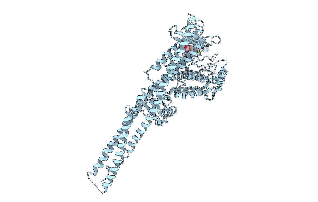
4IWP image
Entry Detail
PDB ID:
4IWP
Title:
Crystal structure and mechanism of activation of TBK1
Biological Source:
Source Organism:
Host Organism:
PDB Version:
Deposition Date:
2013-01-24
Release Date:
2013-03-13
Method Details:
Experimental Method:
Resolution:
3.07 Å
R-Value Free:
0.33
R-Value Work:
0.27
R-Value Observed:
0.28
Space Group:
P 32 2 1
Macromolecular Entities
Polymer Type:polypeptide(L)
Description:Serine/threonine-protein kinase TBK1
Mutations:S172A
Chain IDs:A
Chain Length:659
Number of Molecules:1
Biological Source:Homo sapiens
Ligand Molecules
Primary Citation
Crystal structure and mechanism of activation of TANK-binding kinase 1.
Cell Rep 3 734 746 (2013)
PMID: 23453971 DOI: 10.1016/j.celrep.2013.01.034

Abstact

Tank-binding kinase I (TBK1) plays a key role in the innate immune system by integrating signals from pattern-recognition receptors. Here, we report the X-ray crystal structures of inhibitor-bound inactive and active TBK1 determined to 2.6 Å and 4.0 Å resolution, respectively. The structures reveal a compact dimer made up of trimodular subunits containing an N-terminal kinase domain (KD), a ubiquitin-like domain (ULD), and an α-helical scaffold dimerization domain (SDD). Activation rearranges the KD into an active conformation while maintaining the overall dimer conformation. Low-resolution SAXS studies reveal that the missing C-terminal domain (CTD) extends away from the main body of the kinase dimer. Mutants that interfere with TBK1 dimerization show significantly reduced trans-autophosphorylation but retain the ability to bind adaptor proteins through the CTD. Our results provide detailed insights into the architecture of TBK1 and the molecular mechanism of activation.

Legend

Protein

Chemical

Disease

Primary Citation of related structures