Deposition Date
2013-01-18
Release Date
2013-03-27
Last Version Date
2023-09-20
Entry Detail
PDB ID:
4ITU
Keywords:
Title:
Crystal structure of S-2-HYDROXYPROPYL COENZYME M DEHYDROGENASE (S-HPCDH) bound to S-HPC AND NADH
Biological Source:
Source Organism(s):
Xanthobacter autotrophicus (Taxon ID: 78245)
Expression System(s):
Method Details:
Experimental Method:
Resolution:
1.60 Å
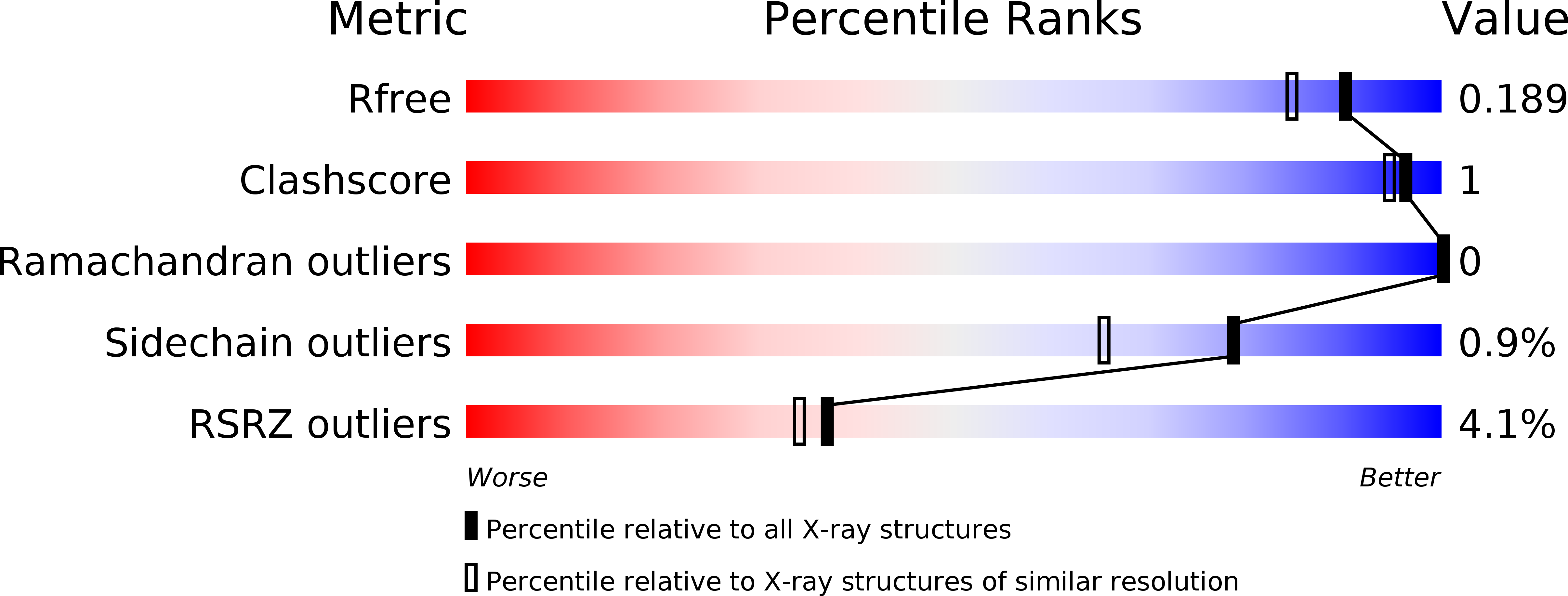
R-Value Free:
0.19
R-Value Work:
0.16
R-Value Observed:
0.16
Space Group:
P 21 21 2