4IRZ image
Deposition Date 2013-01-15
Release Date 2013-09-25
Last Version Date 2024-10-30
Entry Detail
PDB ID:
4IRZ
Keywords:
Title:
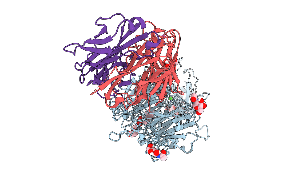
Crystal structure of A4b7 headpiece complexed with Fab Natalizumab
Biological Source:
Source Organism:
Host Organism:
Method Details:
Experimental Method:
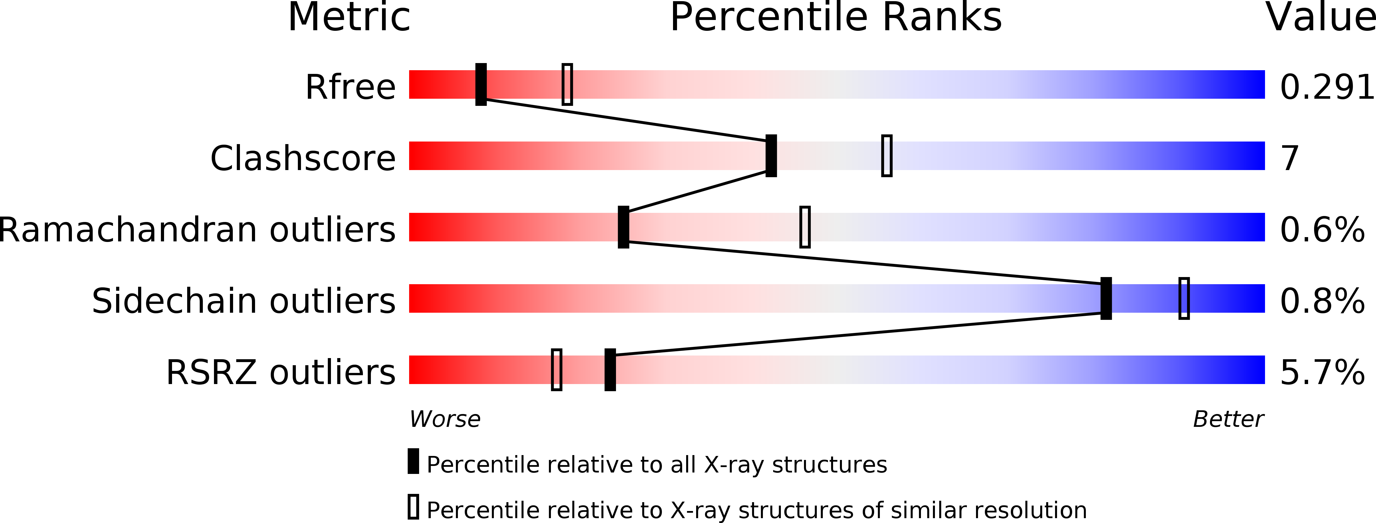
Resolution:
2.84 Å
R-Value Free:
0.28
R-Value Work:
0.23
R-Value Observed:
0.23
Space Group:
P 21 21 21
Macromolecular Entities
Polymer Type:polypeptide(L)
Molecule:Integrin alpha4 subunit
Chain IDs:A
Chain Length:597
Number of Molecules:1
Biological Source:Oryctolagus cuniculus
Polymer Type:polypeptide(L)
Molecule:Fab Natalizumab heavy chain
Chain IDs:C (auth: H)
Chain Length:222
Number of Molecules:1
Biological Source:Homo sapiens
Polymer Type:polypeptide(L)
Molecule:Fab Natalizumab light chain
Chain IDs:B (auth: L)
Chain Length:210
Number of Molecules:1
Biological Source:Homo sapiens
Modified Residue
Compound ID Chain ID Parent Comp ID Details 2D Image
ASN A ASN GLYCOSYLATION SITE
Primary Citation
How natalizumab binds and antagonizes alpha 4 integrins.
J.Biol.Chem. 288 32314 32325 (2013)
PMID: 24047894 DOI: 10.1074/jbc.M113.501668

Abstact

Natalizumab antibody to α4-integrins is used in therapy of multiple sclerosis and Crohn's disease. A crystal structure of the Fab bound to an α4 integrin β-propeller and thigh domain fragment shows that natalizumab recognizes human-mouse differences on the circumference of the β-propeller domain. The epitope is adjacent to but outside of a ligand-binding groove formed at the interface with the β-subunit βI domain and shows no difference in structure when bound to Fab. Competition between Fab and the ligand vascular cell adhesion molecule (VCAM) for binding to cell surface α4β1 shows noncompetitive antagonism. In agreement, VCAM docking models suggest that binding of domain 1 of VCAM to α4-integrins is unimpeded by the Fab, and that bound Fab requires a change in orientation between domains 1 and 2 of VCAM for binding to α4β1. Mapping of species-specific differences onto α4β1 and α4β7 shows that their ligand-binding sites are highly conserved. Skewing away from these conserved regions of the epitopes recognized by current therapeutic function-blocking antibodies has resulted in previously unanticipated mechanisms of action.

Legend

Protein

Chemical

Disease

Primary Citation of related structures