4IRV image
Deposition Date 2013-01-15
Release Date 2014-01-15
Last Version Date 2024-10-16
Entry Detail
PDB ID:
4IRV
Keywords:
Title:
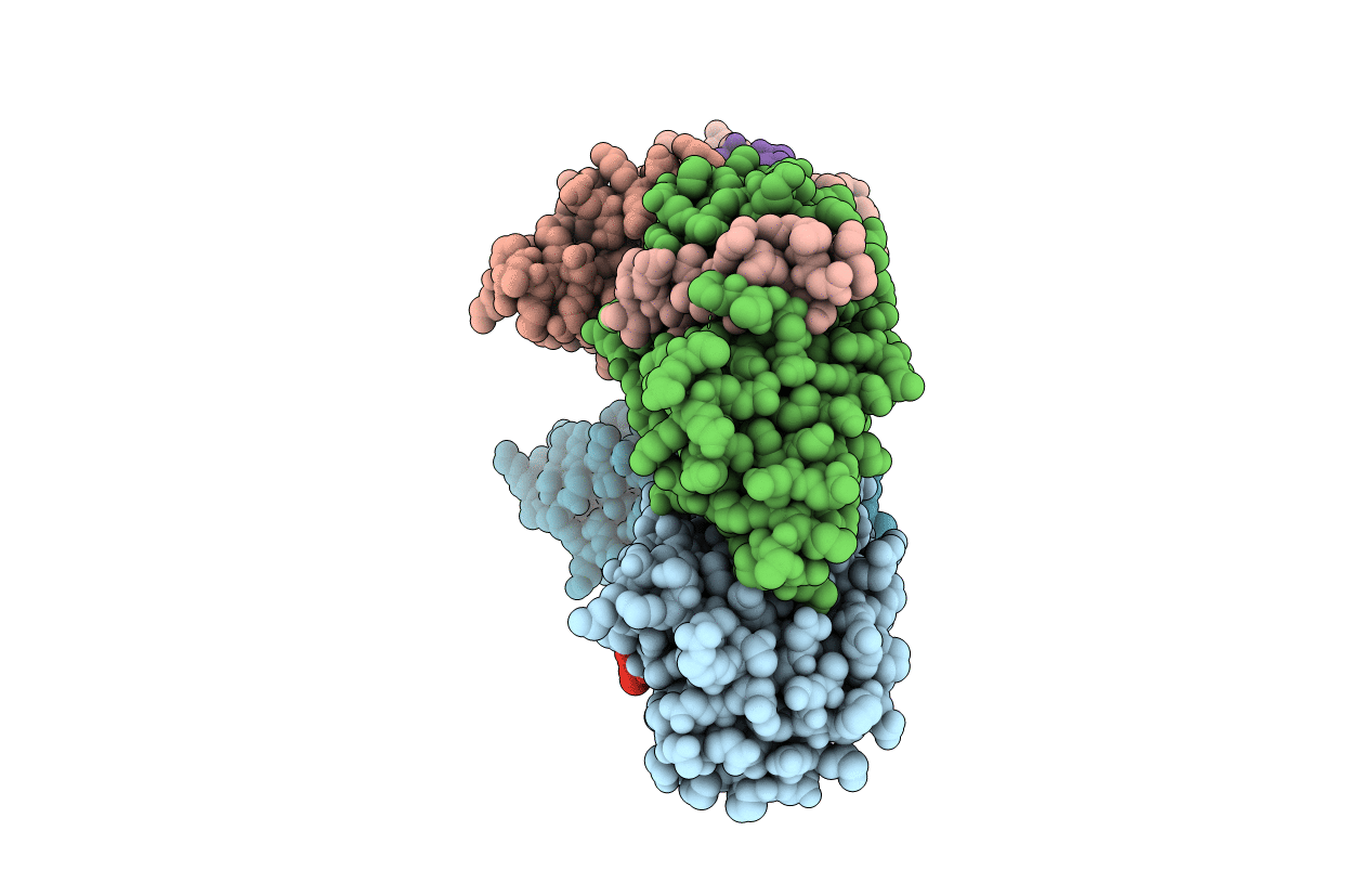
Structure of the Helicobacter pylori CagA Oncogene Bound to the Human Tumor Suppressor Apoptosis-stimulating Protein of p53-2
Biological Source:
Source Organism:
Host Organism:
Method Details:
Experimental Method:
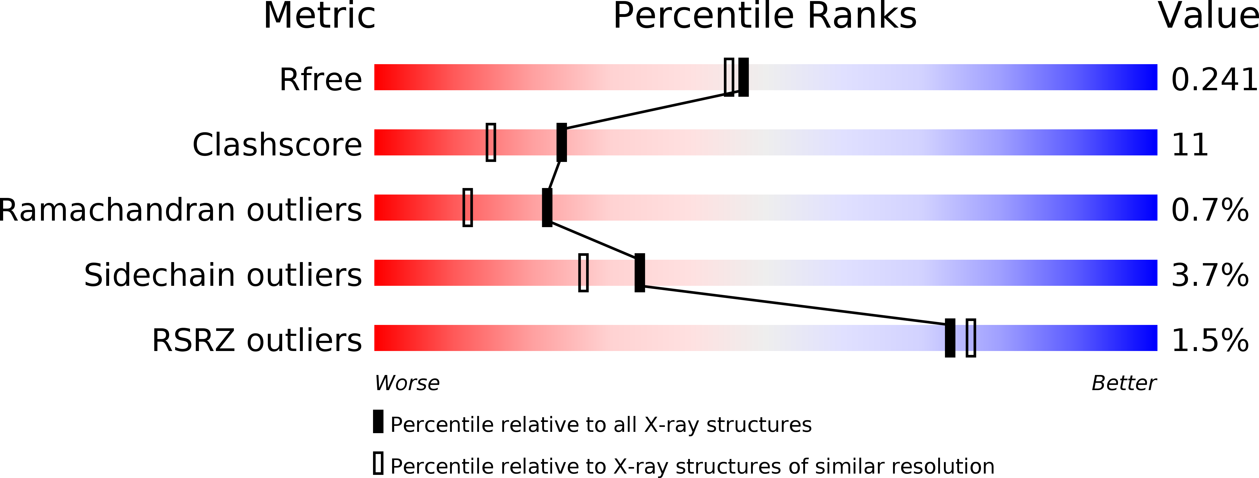
Resolution:
2.04 Å
R-Value Free:
0.23
R-Value Work:
0.18
R-Value Observed:
0.19
Space Group:
C 1 2 1
Macromolecular Entities
Structures with similar UniProt ID
Protein Blast
Polymer Type:polypeptide(L)
Molecule:Cytotoxicity-associated immunodominant antigen
Gene (Uniprot):cagA
Chain IDs:A, B, C, D
Chain Length:221
Number of Molecules:4
Biological Source:Helicobacter pylori
Structures with similar UniProt ID
Protein Blast
Polymer Type:polypeptide(L)
Molecule:Apoptosis-stimulating of p53 protein 2
Gene (Uniprot):TP53BP2
Chain IDs:E, F, G, H
Chain Length:62
Number of Molecules:4
Biological Source:Homo sapiens
Modified Residue
Compound ID Chain ID Parent Comp ID Details 2D Image
MSE A MET SELENOMETHIONINE
Primary Citation
Structure of the Helicobacter pylori CagA oncoprotein bound to the human tumor suppressor ASPP2.
Proc.Natl.Acad.Sci.USA 111 1562 1567 (2014)
PMID: 24474782 DOI: 10.1073/pnas.1320631111

Abstact

The Cytotoxin associated gene A (CagA) protein of Helicobacter pylori is associated with increased virulence and risk of cancer. Recent proteomic studies have demonstrated an association of CagA with the human tumor suppressor Apoptosis-stimulating Protein of p53-2 (ASPP2). We present here a genetic, biochemical, and structural analysis of CagA with ASPP2. Domain delineation of the 120-kDa CagA protein revealed a stable N-terminal subdomain that was used in a yeast two-hybrid screen that identified the proline-rich domain of ASPP2 as a host cellular target. Biochemical experiments confirm this interaction. The cocrystal structure to 2.0-Å resolution of this N-terminal subdomain of CagA with a 7-kDa proline-rich sequence of ASPP2 reveals that this domain of CagA forms a highly specialized three-helix bundle, with large insertions in the loops connecting the helices. These insertions come together to form a deep binding cleft for a highly conserved 20-aa peptide of ASPP2. ASPP2 forms an extended helix in this groove of CagA, burying more than 1,000 Å(2) of surface area. This interaction is disrupted in vitro and in vivo by structure-based, loss-of-contact point mutations of key residues in either CagA or ASPP2. Disruption of CagA and ASPP2 binding alters the function of ASPP2 and leads to the decreased survival of H. pylori-infected cells.

Legend

Protein

Chemical

Disease

Primary Citation of related structures
Feedback Form
Name
Email
Institute
Feedback