4IQH image
Deposition Date 2013-01-11
Release Date 2013-12-18
Last Version Date 2023-09-20
Entry Detail
PDB ID:
4IQH
Title:
Crystal Structure Analysis of Dysferlin C2A variant 1 (C2Av1)
Biological Source:
Source Organism:
Homo sapiens (Taxon ID: 9606)
Host Organism:
Method Details:
Experimental Method:
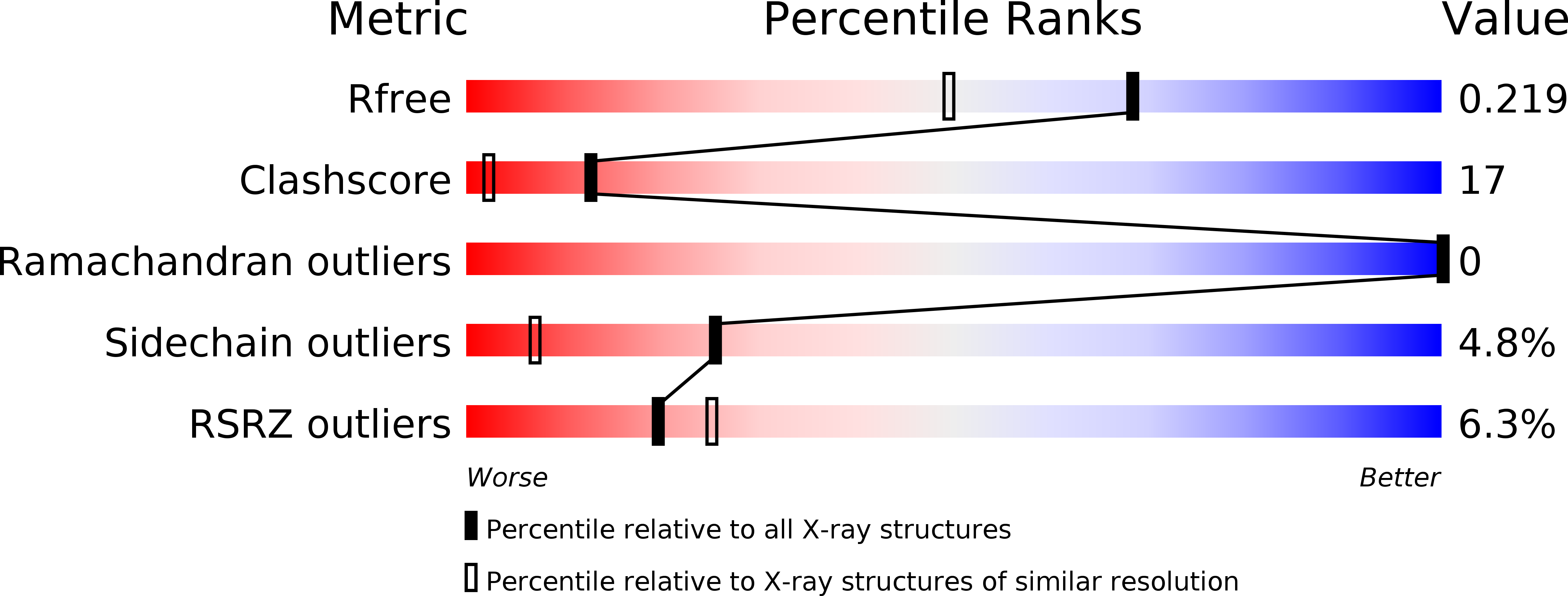
Resolution:
1.76 Å
R-Value Free:
0.21
R-Value Work:
0.19
R-Value Observed:
0.19
Space Group:
P 31 2 1
Macromolecular Entities
Structures with similar UniProt ID
Protein Blast
Polymer Type:polypeptide(L)
Molecule:Dysferlin
Gene (Uniprot):DYSF
Mutagens:C3A
Chain IDs:A, B, C
Chain Length:130
Number of Molecules:3
Biological Source:Homo sapiens
Primary Citation
Alternate Splicing of Dysferlin C2A Confers Ca(2+)-Dependent and Ca(2+)-Independent Binding for Membrane Repair.
Structure 22 104 115 (2014)
PMID: 24239457 DOI: 10.1016/j.str.2013.10.001

Abstact

Dysferlin plays a critical role in the Ca²⁺-dependent repair of microlesions that occur in the muscle sarcolemma. Of the seven C2 domains in dysferlin, only C2A is reported to bind both Ca²⁺ and phospholipid, thus acting as a key sensor in membrane repair. Dysferlin C2A exists as two isoforms, the "canonical" C2A and C2A variant 1 (C2Av1). Interestingly, these isoforms have markedly different responses to Ca²⁺ and phospholipid. Structural and thermodynamic analyses are consistent with the canonical C2A domain as a Ca²⁺-dependent, phospholipid-binding domain, whereas C2Av1 would likely be Ca²⁺-independent under physiological conditions. Additionally, both isoforms display remarkably low free energies of stability, indicative of a highly flexible structure. The inverted ligand preference and flexibility for both C2A isoforms suggest the capability for both constitutive and Ca²⁺-regulated effector interactions, an activity that would be essential in its role as a mediator of membrane repair.

Legend

Protein

Chemical

Disease

Primary Citation of related structures