4IOU image
Deposition Date 2013-01-08
Release Date 2013-05-29
Last Version Date 2023-09-20
Entry Detail
PDB ID:
4IOU
Keywords:
Title:
Crystal structure of the HIV-1 Vif binding, catalytically active domain of APOBEC3F
Biological Source:
Source Organism:
Homo sapiens (Taxon ID: 9606)
Host Organism:
Method Details:
Experimental Method:
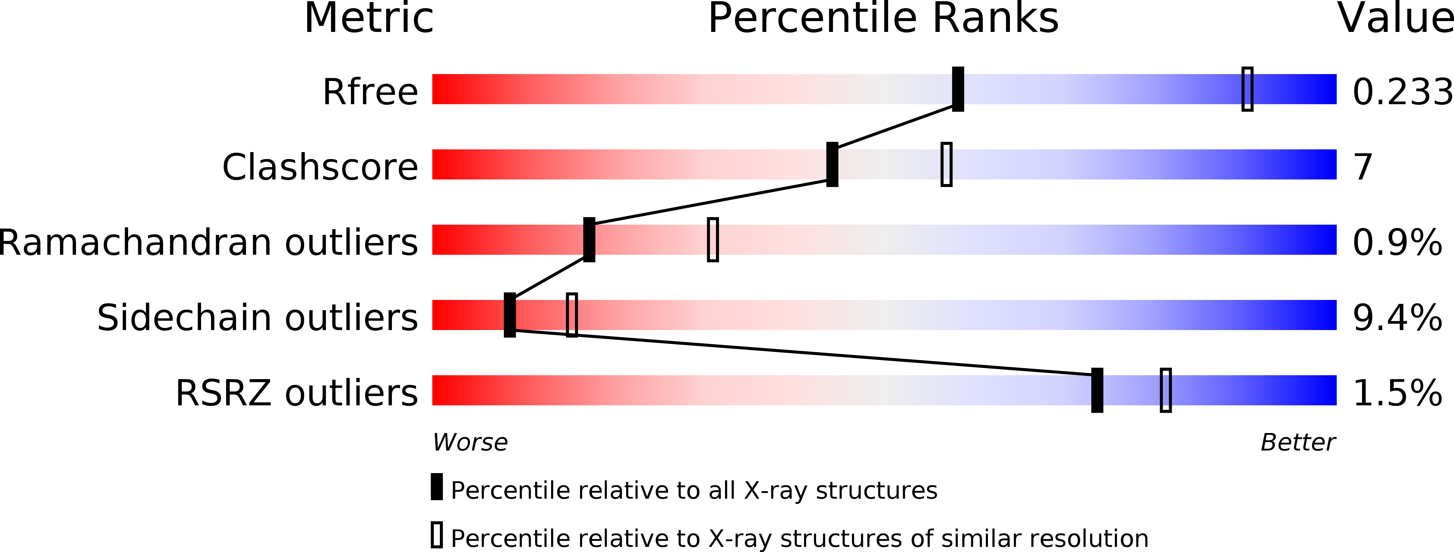
Resolution:
2.75 Å
R-Value Free:
0.23
R-Value Work:
0.19
R-Value Observed:
0.19
Space Group:
P 1
Macromolecular Entities
Polymer Type:polypeptide(L)
Molecule:DNA dC->dU-editing enzyme APOBEC-3F
Gene (Uniprot):APOBEC3F
Mutations:Y196D, H247G, C248R, C259A, F302D, W310K, Y314Y, Q315A, F363D, K355D, K358D
Chain IDs:A, B, C, D
Chain Length:199
Number of Molecules:4
Biological Source:Homo sapiens
Ligand Molecules
Primary Citation
Crystal Structure of the DNA Cytosine Deaminase APOBEC3F: The Catalytically Active and HIV-1 Vif-Binding Domain.
Structure 21 1042 1050 (2013)
PMID: 23685212 DOI: 10.1016/j.str.2013.04.010

Abstact

Human APOBEC3F is an antiretroviral single-strand DNA cytosine deaminase, susceptible to degradation by the HIV-1 protein Vif. In this study the crystal structure of the HIV Vif binding, catalytically active, C-terminal domain of APOBEC3F (A3F-CTD) was determined. The A3F-CTD shares structural motifs with portions of APOBEC3G-CTD, APOBEC3C, and APOBEC2. Residues identified to be critical for Vif-dependent degradation of APOBEC3F all fit within a predominantly negatively charged contiguous region on the surface of A3F-CTD. Specific sequence motifs, previously shown to play a role in Vif susceptibility and virion encapsidation, are conserved across APOBEC3s and between APOBEC3s and HIV-1 Vif. In this structure these motifs pack against each other at intermolecular interfaces, providing potential insights both into APOBEC3 oligomerization and Vif interactions.

Legend

Protein

Chemical

Disease

Primary Citation of related structures