4IO4 image
Deposition Date 2013-01-07
Release Date 2013-02-20
Last Version Date 2024-11-20
Entry Detail
PDB ID:
4IO4
Title:
Crystal Structure of the AvGluR1 ligand binding domain complex with serine at 1.94 Angstrom resolution
Biological Source:
Source Organism:
Adineta vaga (Taxon ID: 104782)
Host Organism:
Method Details:
Experimental Method:
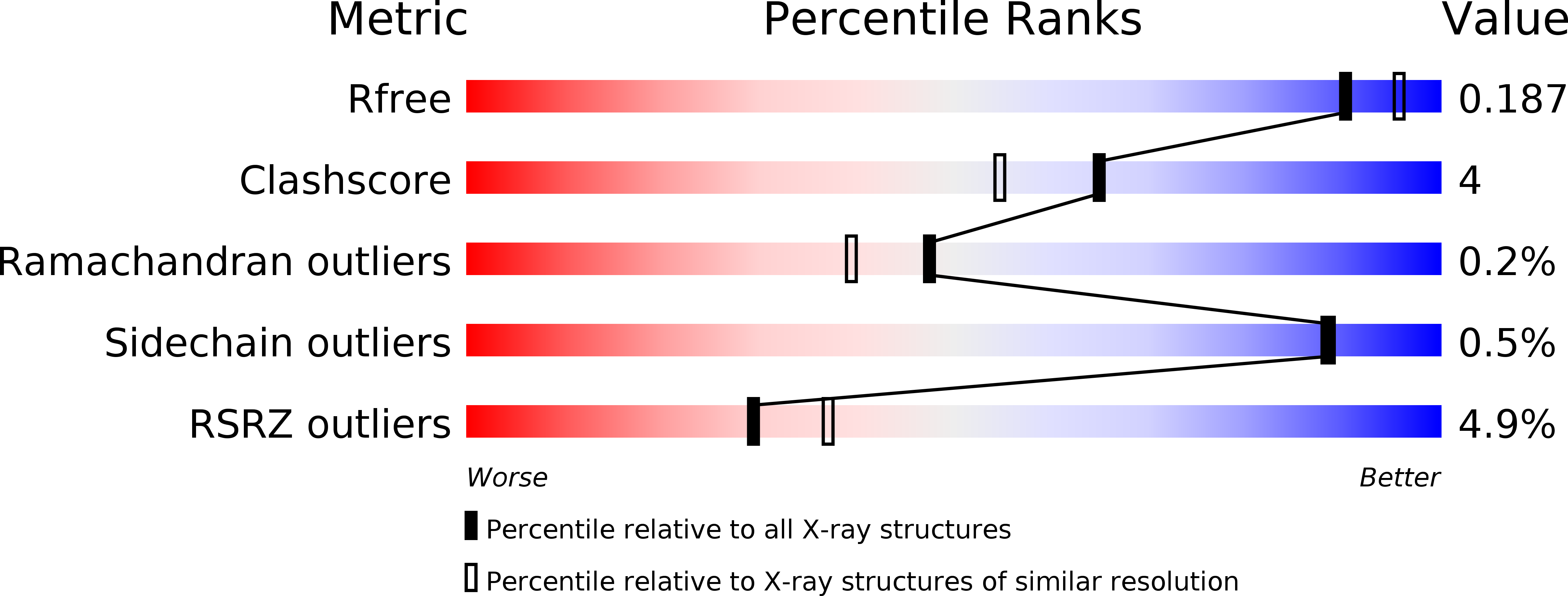
Resolution:
1.94 Å
R-Value Free:
0.18
R-Value Work:
0.14
R-Value Observed:
0.14
Space Group:
P 1 21 1
Macromolecular Entities
Structures with similar UniProt ID
Protein Blast
Polymer Type:polypeptide(L)
Molecule:AvGluR1 ligand binding domain
Chain IDs:A, B
Chain Length:248
Number of Molecules:2
Biological Source:Adineta vaga
Primary Citation
Anions Mediate Ligand Binding in Adineta vaga Glutamate Receptor Ion Channels.
Structure 21 414 425 (2013)
PMID: 23434404 DOI: 10.1016/j.str.2013.01.006

Abstact

AvGluR1, a glutamate receptor ion channel from the primitive eukaryote Adineta vaga, is activated by alanine, cysteine, methionine, and phenylalanine, which produce lectin-sensitive desensitizing responses like those to glutamate, aspartate, and serine. AvGluR1 LBD crystal structures reveal an unusual scheme for binding dissimilar ligands that may be utilized by distantly related odorant/chemosensory receptors. Arginine residues in domain 2 coordinate the γ-carboxyl group of glutamate, whereas in the alanine, methionine, and serine complexes a chloride ion acts as a surrogate ligand, replacing the γ-carboxyl group. Removal of Cl(-) lowers affinity for these ligands but not for glutamate or aspartate nor for phenylalanine, which occludes the anion binding site and binds with low affinity. AvGluR1 LBD crystal structures and sedimentation analysis also provide insights into the evolutionary link between prokaryotic and eukaryotic iGluRs and reveal features unique to both classes, emphasizing the need for additional structure-based studies on iGluR-ligand interactions.

Legend

Protein

Chemical

Disease

Primary Citation of related structures