Deposition Date
2012-12-29
Release Date
2013-04-03
Last Version Date
2024-02-28
Entry Detail
PDB ID:
4IL7
Keywords:
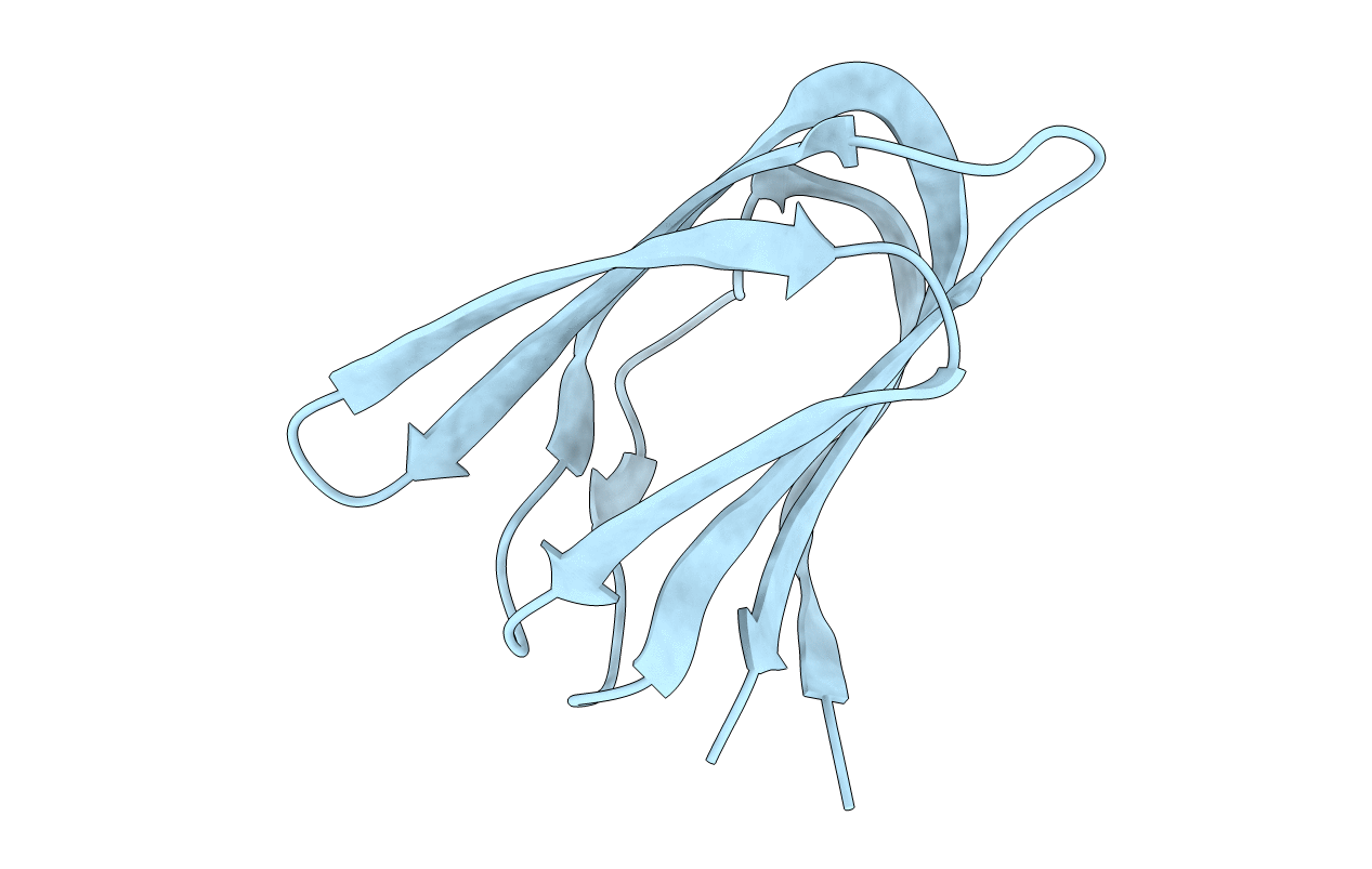
Title:
Crystal structure of A223 C-terminal domain, a structural protein from sulfolobus turreted icosahedral virus (STIV)
Biological Source:
Source Organism(s):
Sulfolobus turreted icosahedral virus (Taxon ID: 269145)
Expression System(s):
Method Details:
Experimental Method:
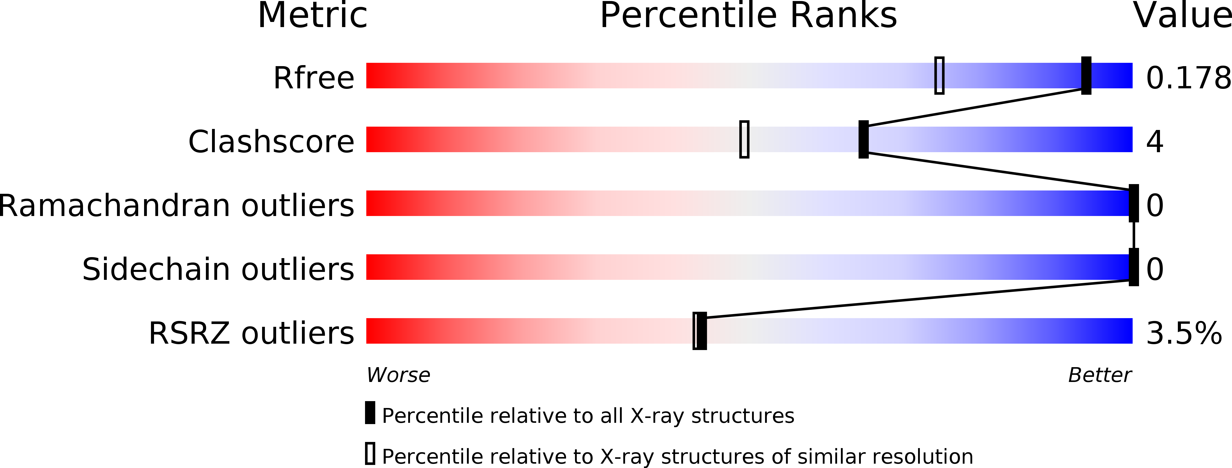
Resolution:
1.40 Å
R-Value Free:
0.16
R-Value Work:
0.14
R-Value Observed:
0.14
Space Group:
P 32 2 1