4IHK image
Deposition Date 2012-12-19
Release Date 2013-12-18
Last Version Date 2023-11-08
Entry Detail
PDB ID:
4IHK
Keywords:
Title:
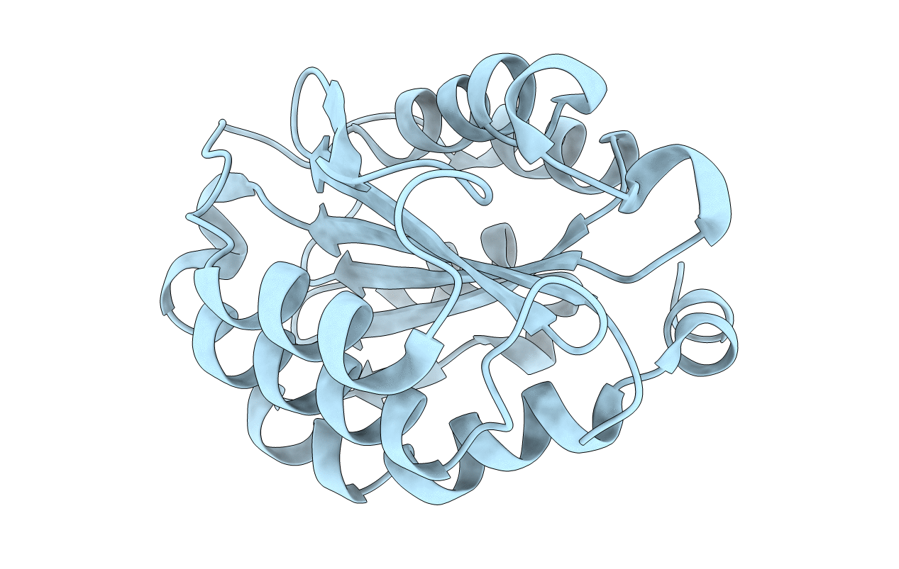
Crystal structure of the Collagen VI alpha3 N5 domain R1061Q
Biological Source:
Source Organism:
Mus musculus (Taxon ID: 10090)
Host Organism:
Method Details:
Experimental Method:
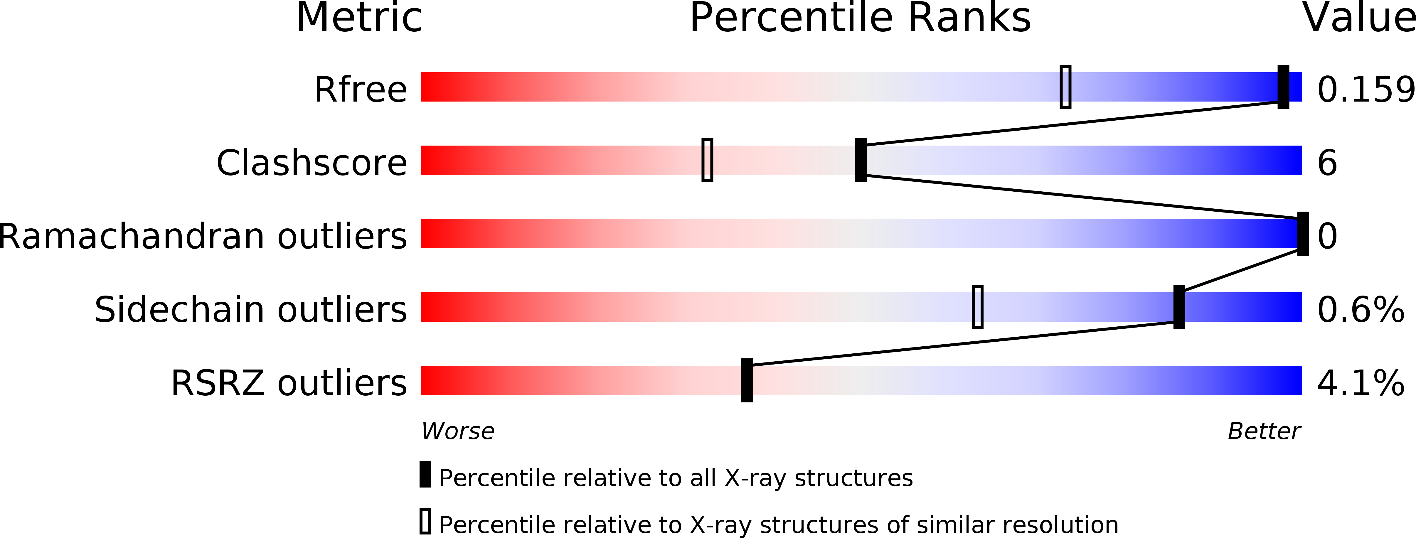
Resolution:
1.20 Å
R-Value Free:
0.16
R-Value Work:
0.13
R-Value Observed:
0.14
Space Group:
P 43 21 2
Macromolecular Entities
Structures with similar UniProt ID
Protein Blast
Polymer Type:polypeptide(L)
Molecule:Collagen alpha3(VI)
Mutagens:G1061R
Chain IDs:A
Chain Length:203
Number of Molecules:1
Biological Source:Mus musculus
Primary Citation
A structure of a collagen VI VWA domain displays N and C termini at opposite sides of the protein
Structure 22 199 208 (2014)
PMID: 24332716 DOI: 10.1016/j.str.2013.06.028

Abstact

Von Willebrand factor A (VWA) domains are versatile protein interaction domains with N and C termini in close proximity placing spatial constraints on overall protein structure. The 1.2 Å crystal structures of a collagen VI VWA domain and a disease-causing point mutant show C-terminal extensions that place the N and C termini at opposite ends. This allows a "beads-on-a-string" arrangement of multiple VWA domains as observed for ten N-terminal domains of the collagen VI α3 chain. The extension is linked to the core domain by a salt bridge and two hydrophobic patches. Comparison of the wild-type and a muscular dystrophy-associated mutant structure identifies a potential perturbation of a protein interaction interface and indeed, the secretion of mutant collagen VI tetramers is affected. Homology modeling is used to locate a number of disease-associated mutations and analyze their structural impact, which will allow mechanistic analysis of collagen-VI-associated muscular dystrophy phenotypes.

Legend

Protein

Chemical

Disease

Primary Citation of related structures