4ICY image
Deposition Date 2012-12-11
Release Date 2013-06-12
Last Version Date 2024-04-03
Entry Detail
PDB ID:
4ICY
Keywords:
Title:
Tracing the Evolution of Angucyclinone Monooxygenases: Structural Determinants for C-12b Hydroxylation and Substrate Inhibition in PgaE
Biological Source:
Source Organism:
Host Organism:
Method Details:
Experimental Method:
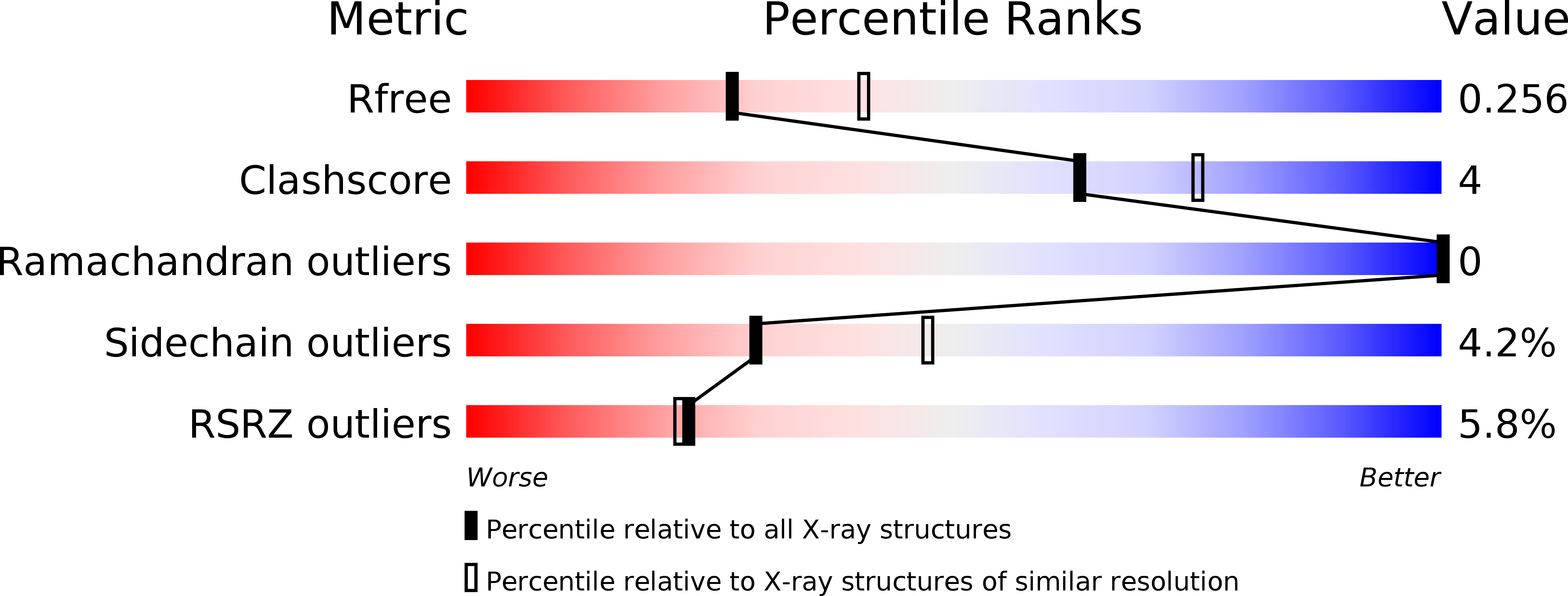
Resolution:
2.40 Å
R-Value Free:
0.25
R-Value Work:
0.19
R-Value Observed:
0.20
Space Group:
F 2 2 2
Macromolecular Entities
Structures with similar UniProt ID
Protein Blast
Polymer Type:polypeptide(L)
Molecule:Polyketide oxygenase PgaE
Mutagens:P78Q, I79F
Chain IDs:A
Chain Length:500
Number of Molecules:1
Biological Source:Streptomyces sp. PGA64
Primary Citation
Tracing the Evolution of Angucyclinone Monooxygenases: Structural Determinants for C-12b Hydroxylation and Substrate Inhibition in PgaE.
Biochemistry 52 4507 4516 (2013)
PMID: 23731237 DOI: 10.1021/bi400381s

Abstact

Two functionally distinct homologous flavoprotein hydroxylases, PgaE and JadH, have been identified as branching points in the biosynthesis of the polyketide antibiotics gaudimycin C and jadomycin A, respectively. These evolutionarily related enzymes are both bifunctional and able to catalyze the same initial reaction, C-12 hydroxylation of the common angucyclinone intermediate prejadomycin. The enzymes diverge in their secondary activities, which include hydroxylation at C-12b by PgaE and dehydration at C-4a/C-12b by JadH. A further difference is that the C-12 hydroxylation is subject to substrate inhibition only in PgaE. Here we have identified regions associated with the C-12b hydroxylation in PgaE by extensive chimeragenesis, focusing on regions surrounding the active site. The results highlight the importance of a hairpin-β motif near the dimer interface, with two nonconserved residues, P78 and I79 (corresponding to Q89 and F90, respectively, in JadH), and invariant residue H73 playing key roles. Kinetic characterization of PgaE variants demonstrates that the secondary C-12b hydroxylation and substrate inhibition by prejadomycin are likely to be interlinked. The crystal structure of the PgaE P78Q/I79F variant at 2.4 Å resolution confirms that the changes do not alter the conformation of the β-strand secondary structure and that the side chains of these residues in effect point away from the active site toward the dimer interface. The results support a catalytic model for PgaE containing two binding modes for C-12 and C-12b hydroxylations, where binding of prejadomycin in the orientation for C-12b hydroxylation leads to substrate inhibition. The presence of an allosteric network is evident based on enzyme kinetics.

Legend

Protein

Chemical

Disease

Primary Citation of related structures