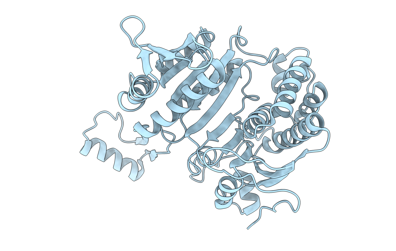
4ICD image
Deposition Date 1989-12-28
Release Date 1991-01-15
Last Version Date 2024-10-09
Entry Detail
PDB ID:
4ICD
Title:
REGULATION OF ISOCITRATE DEHYDROGENASE BY PHOSPHORYLATION INVOLVES NO LONG-RANGE CONFORMATIONAL CHANGE IN THE FREE ENZYME
Biological Source:
Source Organism:
Escherichia coli (Taxon ID: 562)
Method Details:
Experimental Method:
Resolution:
2.50 Å
R-Value Work:
0.16
Space Group:
P 43 21 2
Macromolecular Entities
Structures with similar UniProt ID
Protein Blast
Polymer Type:polypeptide(L)
Molecule:PHOSPHORYLATED ISOCITRATE DEHYDROGENASE
Gene (Uniprot):icd
Chain IDs:A
Chain Length:416
Number of Molecules:1
Biological Source:Escherichia coli
Modified Residue
Compound ID Chain ID Parent Comp ID Details 2D Image
SEP A SER PHOSPHOSERINE
Primary Citation
Regulation of isocitrate dehydrogenase by phosphorylation involves no long-range conformational change in the free enzyme.
J.Biol.Chem. 265 3599 3602 (1990)
PMID: 2406256

Abstact

The structure of the phosphorylated form of isocitrate dehydrogenase from Escherichia coli has been solved and refined to an R-factor of 16.9% at 2.5-A resolution. Comparison with the structure of the dephosphorylated enzyme shows that there are no large scale conformational changes and that small conformational changes are highly localized around the site of phosphorylation at serine 113. Tyrosine 160 rotates by 15 degrees, and there is a local rearrangement of water structure. There is an 0.2-A net movement of loop 230-234, and side chain shifts of 0.2 A root mean square for isoleucine 159 and lysine 199. The lack of large conformational changes, the observation of a possible isocitrate binding site close to serine 113, and the demonstration that the phosphorylated enzyme is unable to bind isocitrate suggest that this enzyme is inactivated by a direct electrostatic interaction between the substrate and the serine phosphate.

Legend

Protein

Chemical

Disease

Primary Citation of related structures