4I8T image
Deposition Date 2012-12-04
Release Date 2013-09-11
Last Version Date 2023-09-20
Entry Detail
PDB ID:
4I8T
Title:
C.Esp1396I bound to a 19 base pair DNA duplex
Biological Source:
Source Organism:
Host Organism:
Method Details:
Experimental Method:
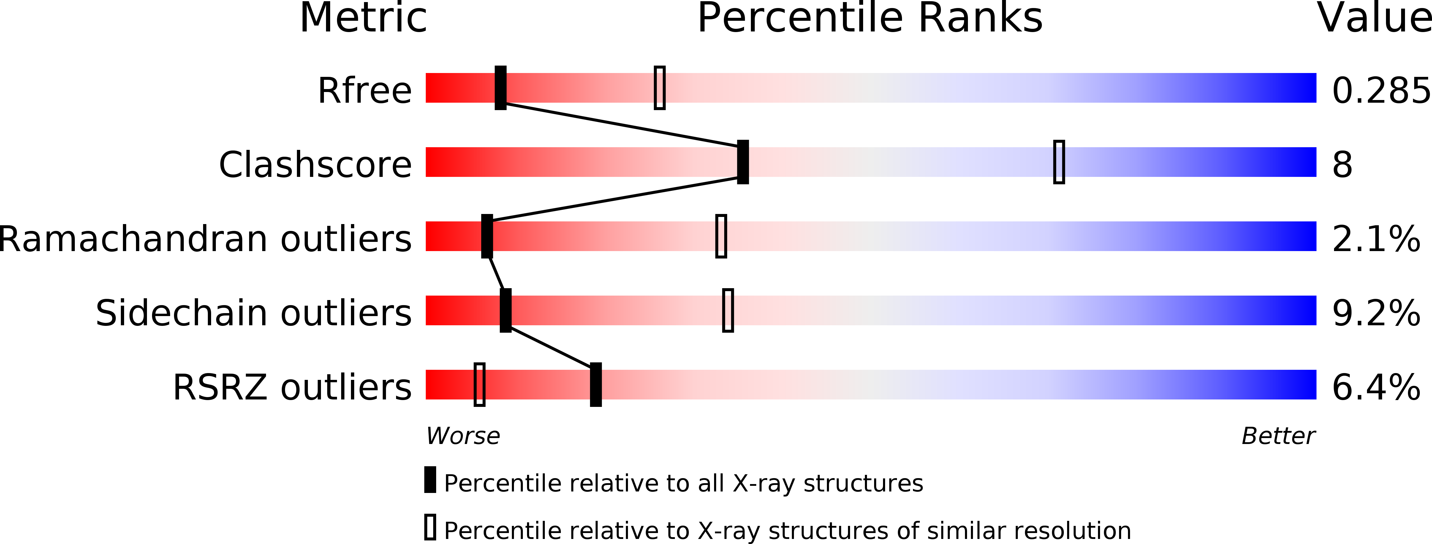
Resolution:
3.00 Å
R-Value Free:
0.29
R-Value Work:
0.23
R-Value Observed:
0.24
Space Group:
C 1 2 1
Macromolecular Entities
Structures with similar UniProt ID
Protein Blast
Polymer Type:polypeptide(L)
Molecule:Regulatory protein
Gene (Uniprot):esp1396IC
Chain IDs:A, B
Chain Length:82
Number of Molecules:2
Biological Source:Enterobacter sp.
Polymer Type:polydeoxyribonucleotide
Molecule:DNA (5'-D(P*TP*GP*TP*TP*GP*AP*CP*TP*AP*TP*AP*AP*TP*CP*AP*CP*AP*CP*A)-3')
Chain IDs:C
Chain Length:19
Number of Molecules:1
Biological Source:
Polymer Type:polydeoxyribonucleotide
Molecule:DNA (5'-D(P*TP*GP*TP*GP*TP*GP*AP*TP*TP*AP*TP*AP*GP*TP*CP*AP*AP*CP*A)-3')
Chain IDs:D
Chain Length:19
Number of Molecules:1
Biological Source:
Ligand Molecules
Primary Citation
Structural analysis of DNA-protein complexes regulating the restriction-modification system Esp1396I.
Acta Crystallogr.,Sect.F 69 962 966 (2013)
PMID: 23989141 DOI: 10.1107/S174430911302126X

Abstact

The controller protein of the type II restriction-modification (RM) system Esp1396I binds to three distinct DNA operator sequences upstream of the methyltransferase and endonuclease genes in order to regulate their expression. Previous biophysical and crystallographic studies have shown molecular details of how the controller protein binds to the operator sites with very different affinities. Here, two protein-DNA co-crystal structures containing portions of unbound DNA from native operator sites are reported. The DNA in both complexes shows significant distortion in the region between the conserved symmetric sequences, similar to that of a DNA duplex when bound by the controller protein (C-protein), indicating that the naked DNA has an intrinsic tendency to bend when not bound to the C-protein. Moreover, the width of the major groove of the DNA adjacent to a bound C-protein dimer is observed to be significantly increased, supporting the idea that this DNA distortion contributes to the substantial cooperativity found when a second C-protein dimer binds to the operator to form the tetrameric repression complex.

Legend

Protein

Chemical

Disease

Primary Citation of related structures
Feedback Form
Name
Email
Institute
Feedback