Deposition Date
2012-12-01
Release Date
2013-10-16
Last Version Date
2024-10-09
Entry Detail
PDB ID:
4I7Y
Keywords:
Title:
Crystal Structure of Human Alpha Thrombin in Complex with a 27-mer Aptamer Bound to Exosite II
Biological Source:
Source Organism(s):
SYNTHETIC DNA (Taxon ID: 32630)
Homo sapiens (Taxon ID: 9606)
Homo sapiens (Taxon ID: 9606)
Method Details:
Experimental Method:
Resolution:
2.40 Å
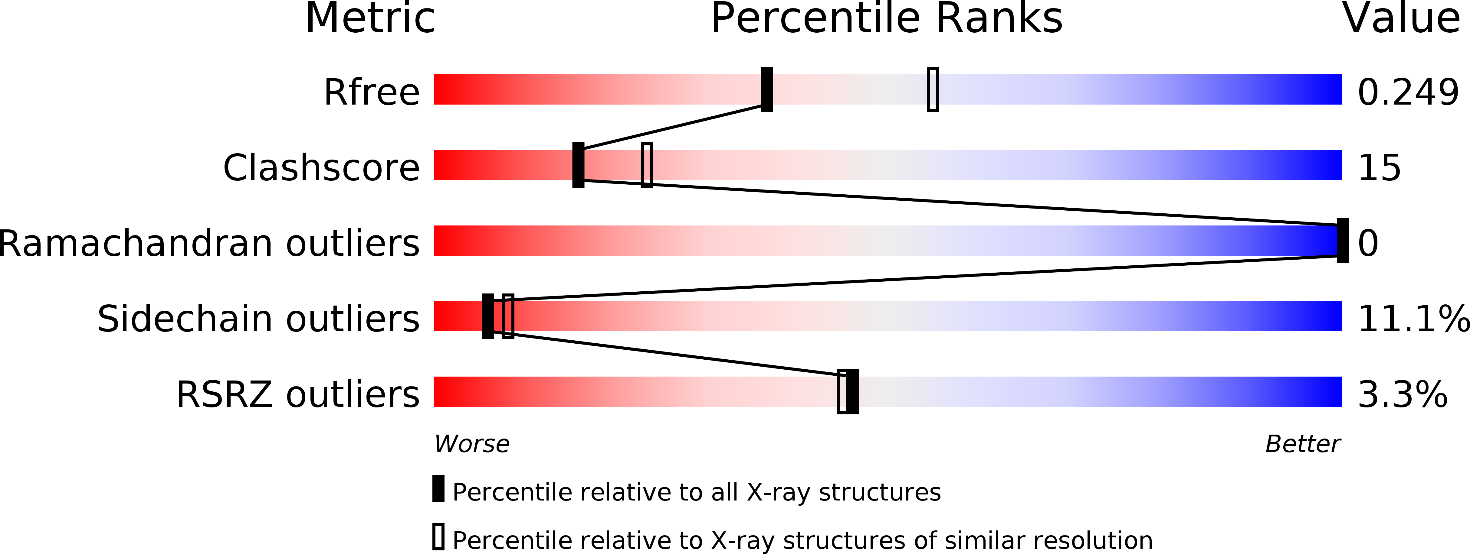
R-Value Free:
0.24
R-Value Work:
0.18
R-Value Observed:
0.18
Space Group:
P 1 21 1