4I6X image
Deposition Date 2012-11-30
Release Date 2013-04-03
Last Version Date 2024-11-06
Entry Detail
PDB ID:
4I6X
Keywords:
Title:
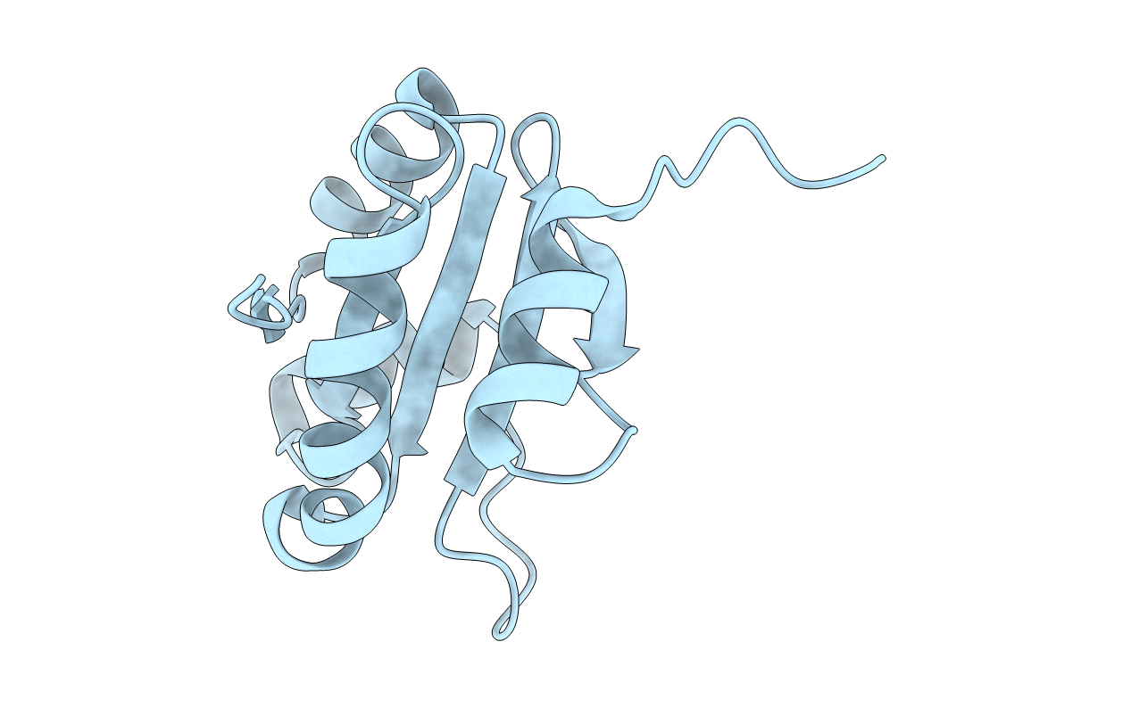
Crystal Structure of Non-catalyic Domain of Protein Disulfide Isomerase-related (PDIr) Protein
Biological Source:
Source Organism:
Homo sapiens (Taxon ID: 9606)
Host Organism:
Method Details:
Experimental Method:
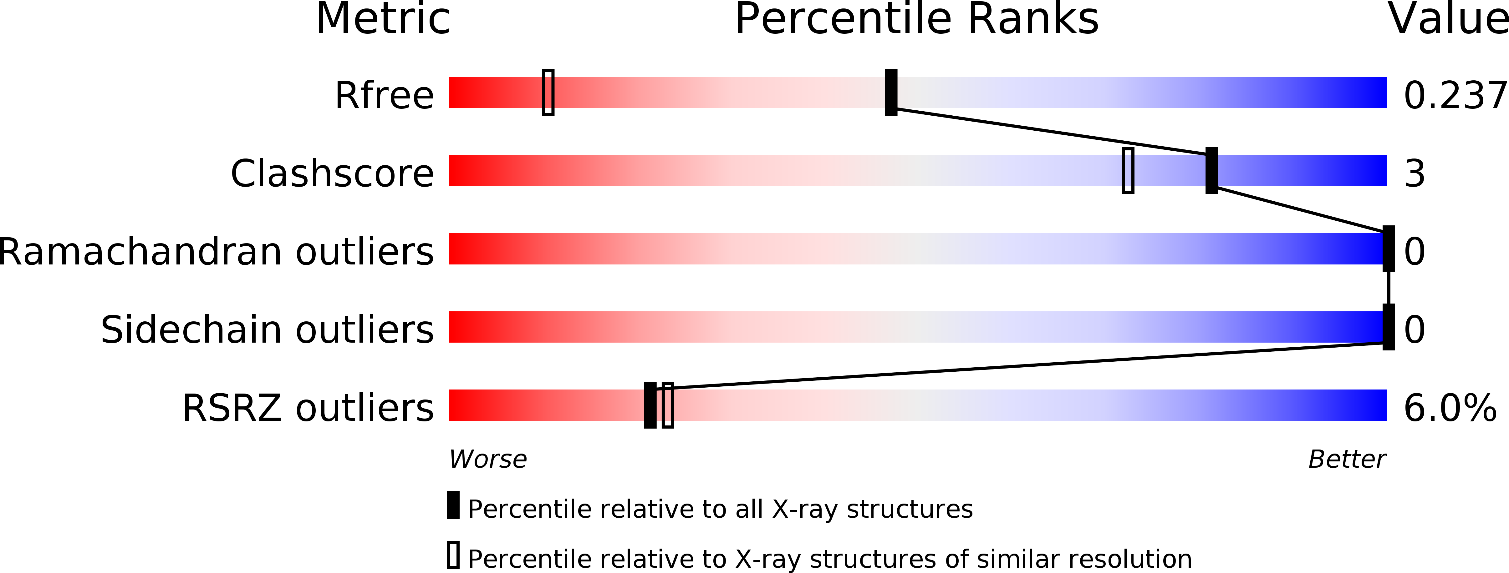
Resolution:
1.50 Å
R-Value Free:
0.23
R-Value Work:
0.19
R-Value Observed:
0.19
Space Group:
P 21 21 21
Macromolecular Entities
Structures with similar UniProt ID
Protein Blast
Polymer Type:polypeptide(L)
Molecule:Protein disulfide-isomerase A5
Gene (Uniprot):PDIA5
Chain IDs:A
Chain Length:131
Number of Molecules:1
Biological Source:Homo sapiens
Primary Citation
Structure of the Non-Catalytic Domain of the Protein Disulfide Isomerase-Related Protein (PDIR) Reveals Function in Protein Binding.
Plos One 8 e62021 e62021 (2013)
PMID: 23614004 DOI: 10.1371/journal.pone.0062021

Abstact

Protein disulfide isomerases comprise a large family of enzymes responsible for catalyzing the proper oxidation and folding of newly synthesized proteins in the endoplasmic reticulum (ER). Protein disulfide isomerase-related (PDIR) protein (also known as PDIA5) is a specialized member that participates in the folding of α1-antitrypsin and N-linked glycoproteins. Here, the crystal structure of the non-catalytic domain of PDIR was determined to 1.5 Å resolution. The structure adopts a thioredoxin-like fold stabilized by a structural disulfide bridge with a positively charged binding surface for interactions with the ER chaperones, calreticulin and ERp72. Crystal contacts between molecules potentially mimic the interactions of PDIR with misfolded substrate proteins. The results suggest that the non-catalytic domain of PDIR plays a key role in the recognition of protein partners and substrates.

Legend

Protein

Chemical

Disease

Primary Citation of related structures