Deposition Date
2012-11-29
Release Date
2013-08-14
Last Version Date
2024-11-20
Entry Detail
PDB ID:
4I60
Keywords:
Title:
Crystal structure of avidin - biotinylruthenocene complex
Biological Source:
Source Organism(s):
Gallus gallus (Taxon ID: 9031)
Method Details:
Experimental Method:
Resolution:
2.50 Å
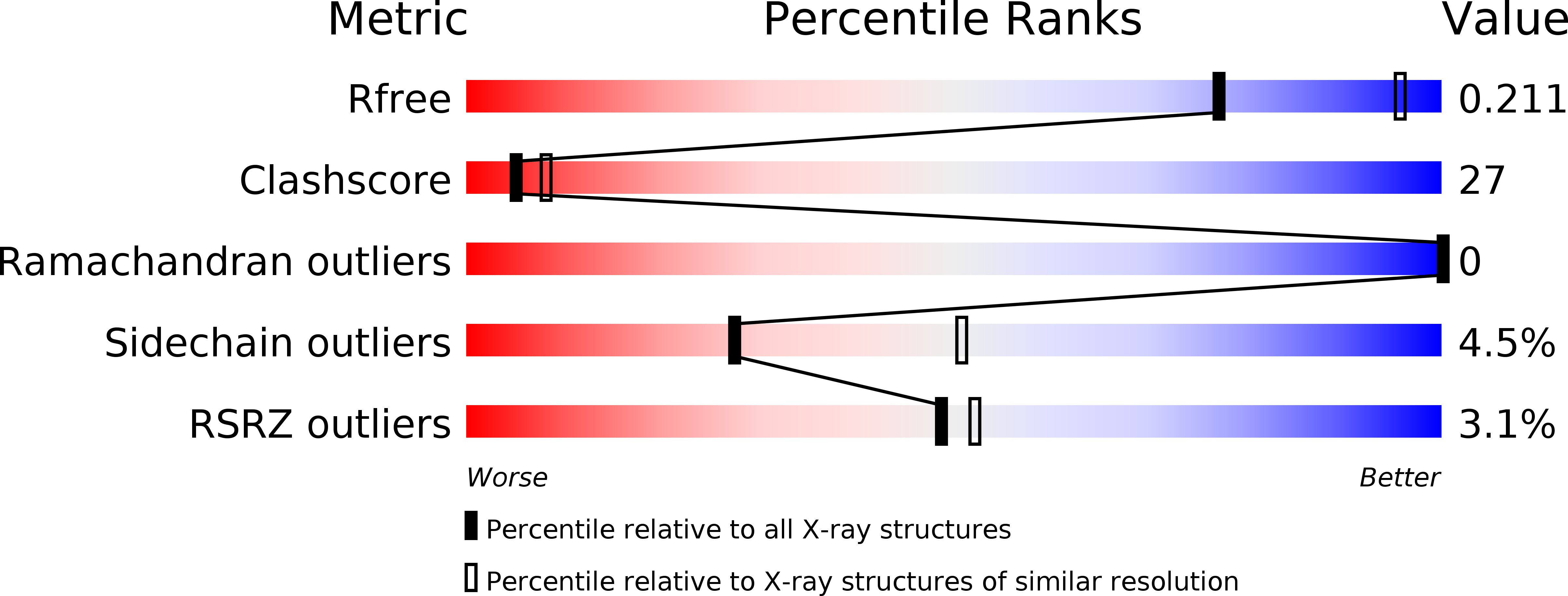
R-Value Free:
0.21
R-Value Work:
0.16
R-Value Observed:
0.16
Space Group:
P 42 21 2