
Deposition Date
2012-11-28
Release Date
2013-12-04
Last Version Date
2024-10-30
Entry Detail
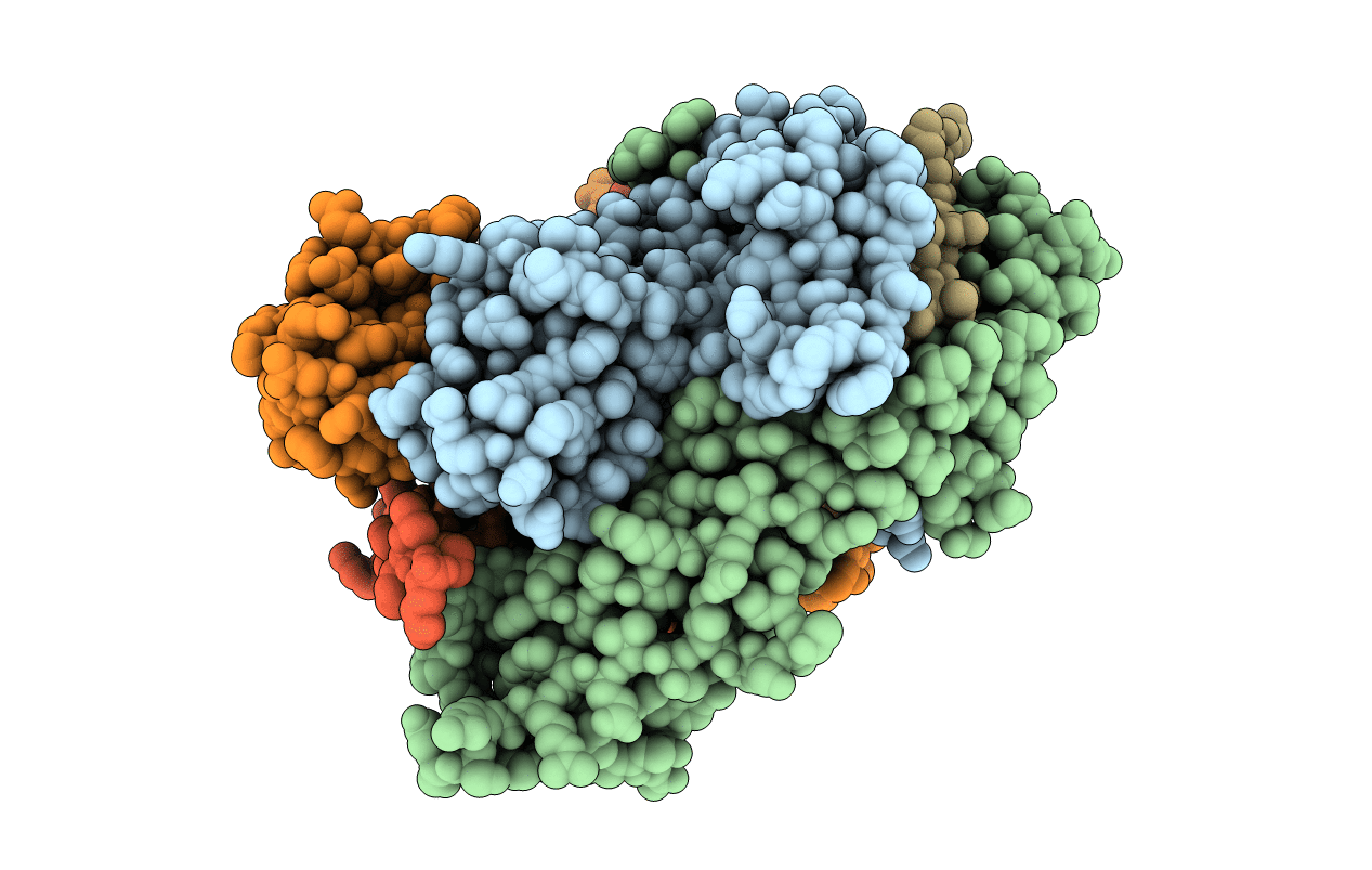
PDB ID:
4I5B
Keywords:
Title:
Structure of human MHC class II protein HLA-DR1 carrying an influenza hemagglutinin peptide partially filling the binding groove
Biological Source:
Source Organism(s):
Homo sapiens (Taxon ID: 9606)
Expression System(s):
Method Details:
Experimental Method:
Resolution:
2.12 Å
R-Value Free:
0.23
R-Value Work:
0.19
R-Value Observed:
0.20
Space Group:
C 2 2 21
