4I3L image
Deposition Date 2012-11-26
Release Date 2013-05-22
Last Version Date 2024-02-28
Entry Detail
PDB ID:
4I3L
Title:
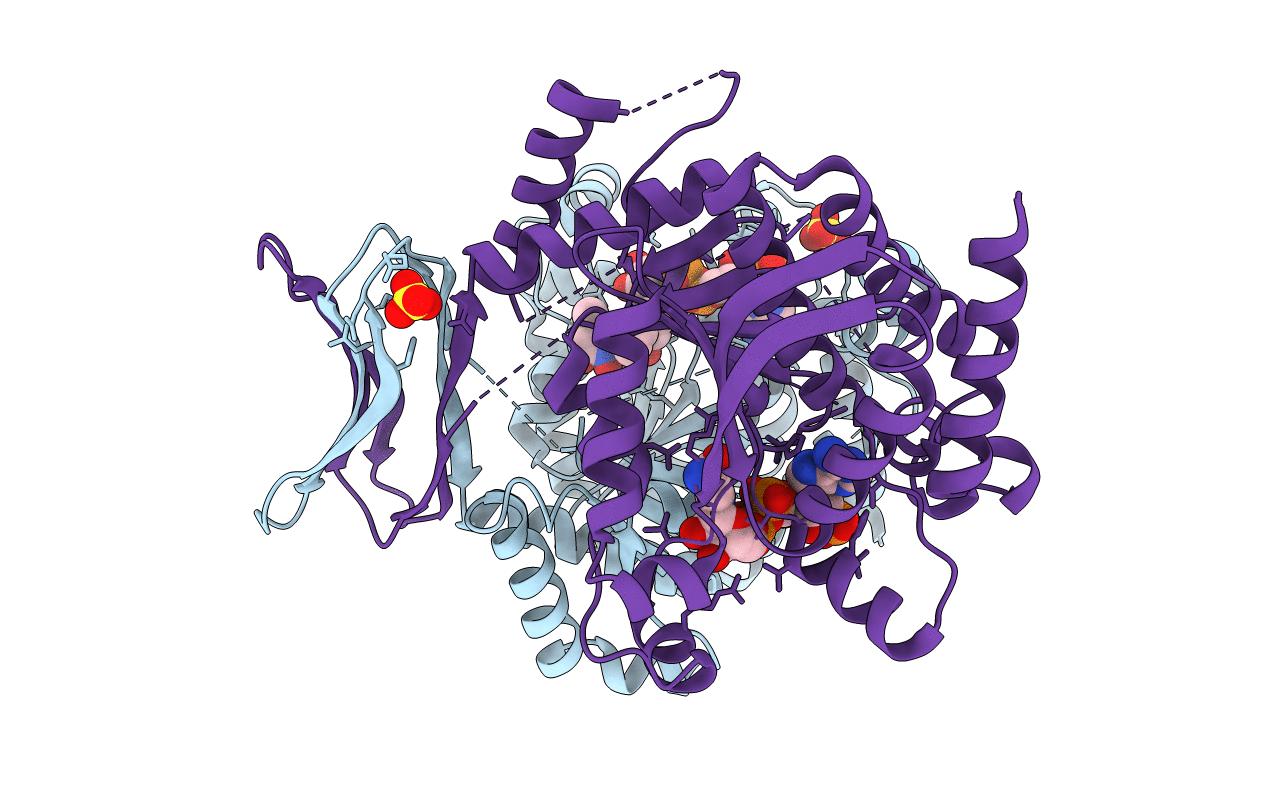
Crystal structure of a metabolic reductase with 6-benzyl-1-hydroxy-4-methylpyridin-2(1H)-one
Biological Source:
Source Organism:
Homo sapiens (Taxon ID: 9606)
Host Organism:
Method Details:
Experimental Method:
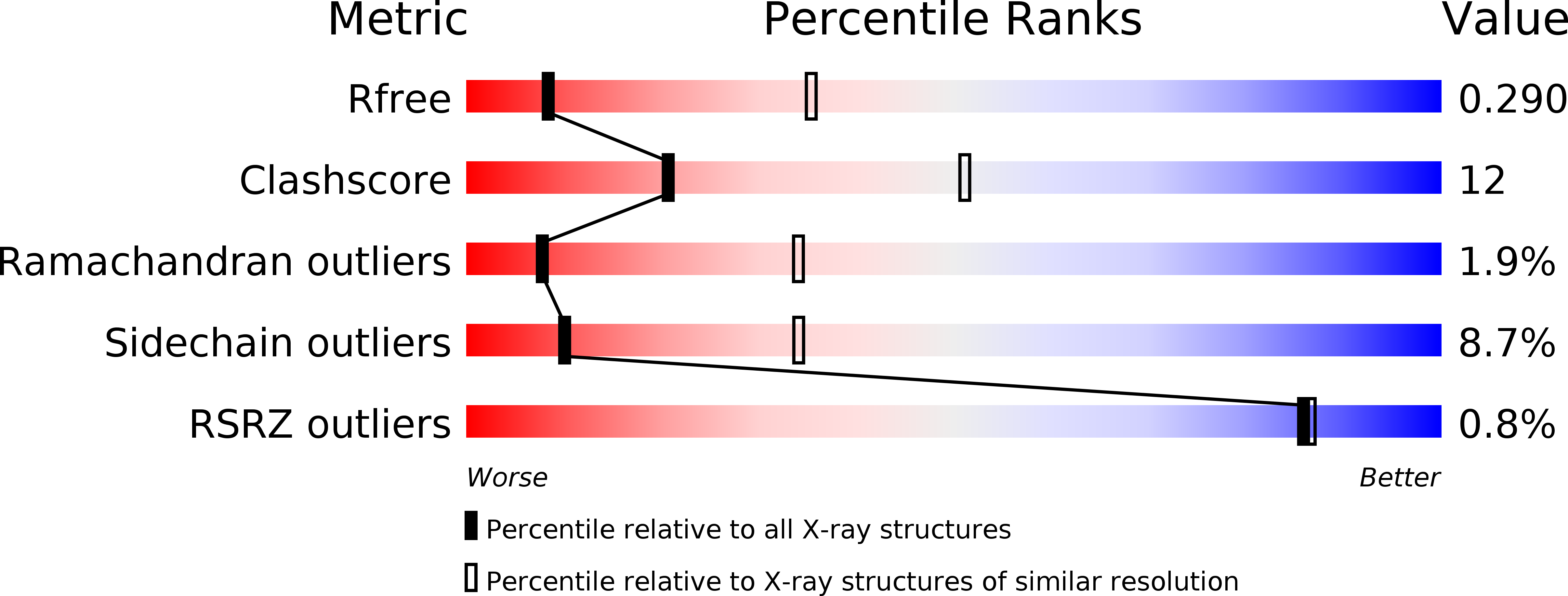
Resolution:
3.29 Å
R-Value Free:
0.29
R-Value Work:
0.21
R-Value Observed:
0.22
Space Group:
P 43 21 2
Macromolecular Entities
Polymer Type:polypeptide(L)
Molecule:Isocitrate dehydrogenase [NADP] cytoplasmic
Gene (Uniprot):IDH1
Mutations:R132H
Chain IDs:A, B
Chain Length:414
Number of Molecules:2
Biological Source:Homo sapiens
Primary Citation
Crystallographic Investigation and Selective Inhibition of Mutant Isocitrate Dehydrogenase.
ACS Med Chem Lett 4 542 546 (2013)
PMID: 23795241 DOI: 10.1021/ml400036z

Abstact

Mutations in isocitrate dehydrogenase (IDH), a key enzyme in the tricarboxylic acid cycle, have recently been found in ~75% glioma and ~20% acute myeloid leukemia. Different from the wild-type enzyme, mutant IDH1 catalyzes the reduction of α-ketoglutaric acid to D-2-hydroxyglutaric acid. Strong evidence has shown mutant IDH1 represents a novel target for this type of cancer. We found two 1-hydroxypyridin-2-one compounds that are potent inhibitors of R132H and R132C IDH1 mutants with Ki values as low as 120 nM. These compounds exhibit >60-fold selectivity against wild-type IDH1 and can inhibit the production of D-2-hydroxyglutaric acid in IDH1 mutated cells, representing novel chemical probes for cancer biology studies. We also report the first inhibitor-bound crystal structures of IDH1(R132H), showing these inhibitors have H-bond, electrostatic and hydrophobic interactions with the mutant enzyme. Comparison with the substrate-bound IDH1 structures revealed the structural basis for the high enzyme selectivity of these compounds.

Legend

Protein

Chemical

Disease

Primary Citation of related structures