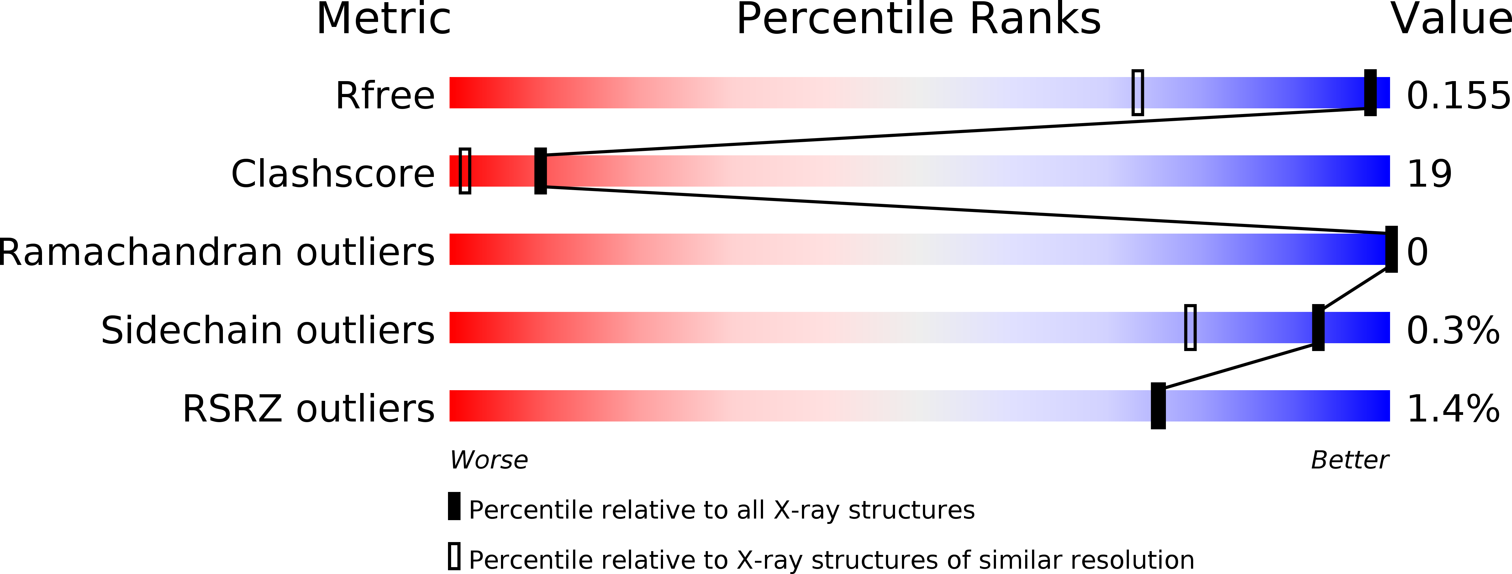
4I3B image
Deposition Date 2012-11-26
Release Date 2013-06-19
Last Version Date 2023-11-08
Entry Detail
PDB ID:
4I3B
Title:
Crystal structure of fluorescent protein UnaG wild type
Biological Source:
Source Organism:
Host Organism:
Method Details:
Experimental Method:
Resolution:
1.20 Å
R-Value Free:
0.15
R-Value Work:
0.12
R-Value Observed:
0.13
Space Group:
P 1 21 1
Macromolecular Entities
Polymer Type:polypeptide(L)
Molecule:Bilirubin-inducible fluorescent protein UnaG
Chain IDs:A, B, C, D, E, F
Chain Length:139
Number of Molecules:6
Biological Source:Anguilla japonica
Primary Citation
A bilirubin-inducible fluorescent protein from eel muscle
Cell(Cambridge,Mass.) 153 1602 1611 (2013)
PMID: 23768684 DOI: 10.1016/j.cell.2013.05.038

Abstact

The fluorescent protein toolbox has revolutionized experimental biology. Despite this advance, no fluorescent proteins have been identified from vertebrates, nor has chromogenic ligand-inducible activation or clinical utility been demonstrated. Here, we report the cloning and characterization of UnaG, a fluorescent protein from Japanese eel. UnaG belongs to the fatty-acid-binding protein (FABP) family, and expression in eel is restricted to small-diameter muscle fibers. On heterologous expression in cell lines or mouse brain, UnaG produces oxygen-independent green fluorescence. Remarkably, UnaG fluorescence is triggered by an endogenous ligand, bilirubin, a membrane-permeable heme metabolite and clinical health biomarker. The holoUnaG structure at 1.2 Å revealed a biplanar coordination of bilirubin by reversible π-conjugation, and we used this high-affinity and high-specificity interaction to establish a fluorescence-based human bilirubin assay with promising clinical utility. UnaG will be the prototype for a versatile class of ligand-activated fluorescent proteins, with applications in research, medicine, and bioengineering.

Legend

Protein

Chemical

Disease

Primary Citation of related structures