Deposition Date
2012-11-23
Release Date
2013-03-13
Last Version Date
2024-11-20
Entry Detail
PDB ID:
4I2Z
Keywords:
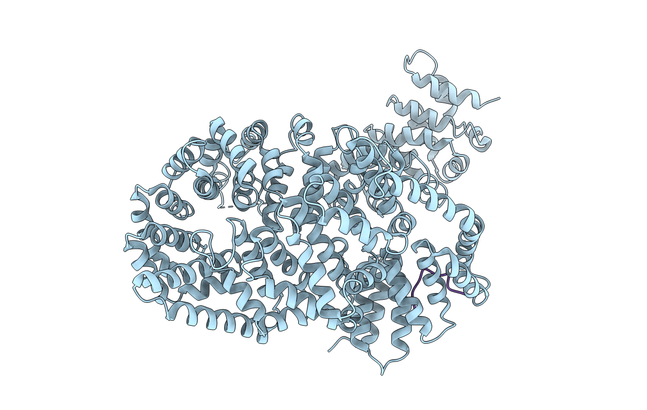
Title:
Crystal structure of the myosin chaperone UNC-45 from C.elegans in complex with a Hsp90 peptide
Biological Source:
Source Organism(s):
Caenorhabditis elegans (Taxon ID: 6239)
Expression System(s):
Method Details:
Experimental Method:
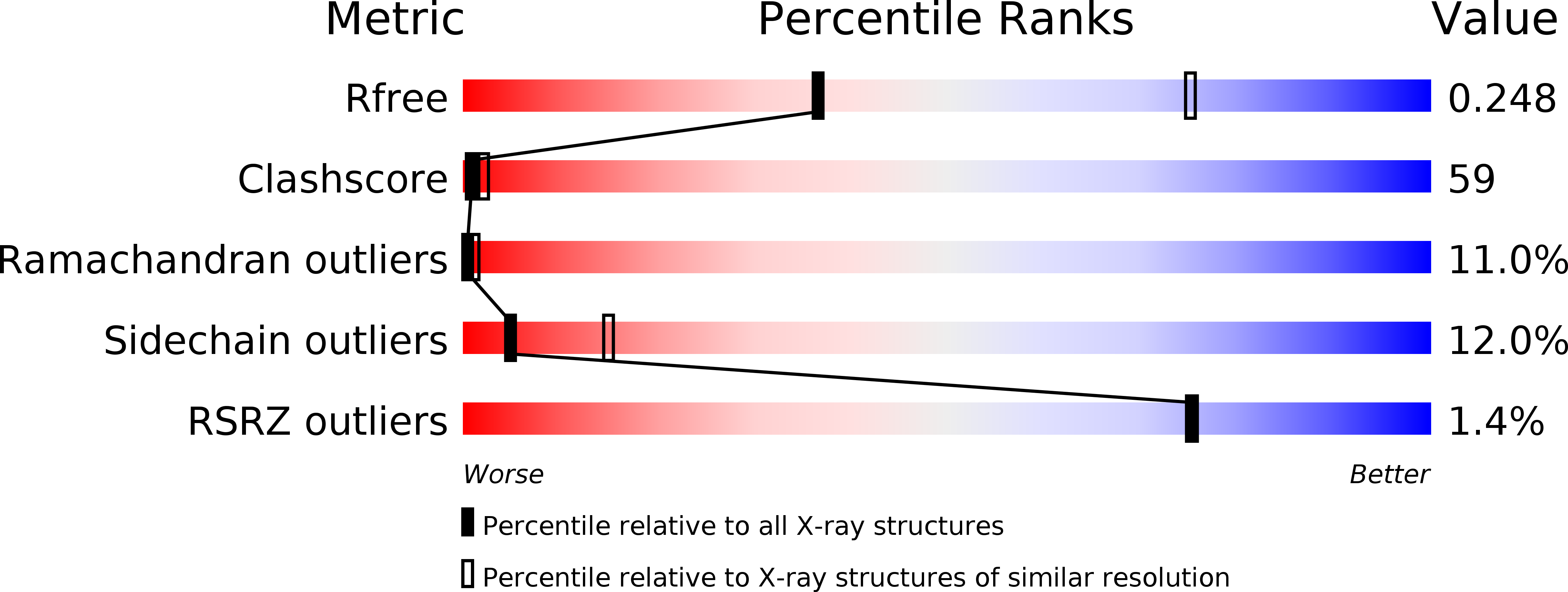
Resolution:
2.90 Å
R-Value Free:
0.25
R-Value Work:
0.23
R-Value Observed:
0.23
Space Group:
P 61 2 2