4I2X image
Deposition Date 2012-11-23
Release Date 2013-12-18
Last Version Date 2024-11-20
Entry Detail
PDB ID:
4I2X
Keywords:
Title:
Crystal structure of Signal Regulatory Protein gamma (SIRP-gamma) in complex with FabOX117
Biological Source:
Source Organism:
Homo sapiens (Taxon ID: 9606)
Method Details:
Experimental Method:
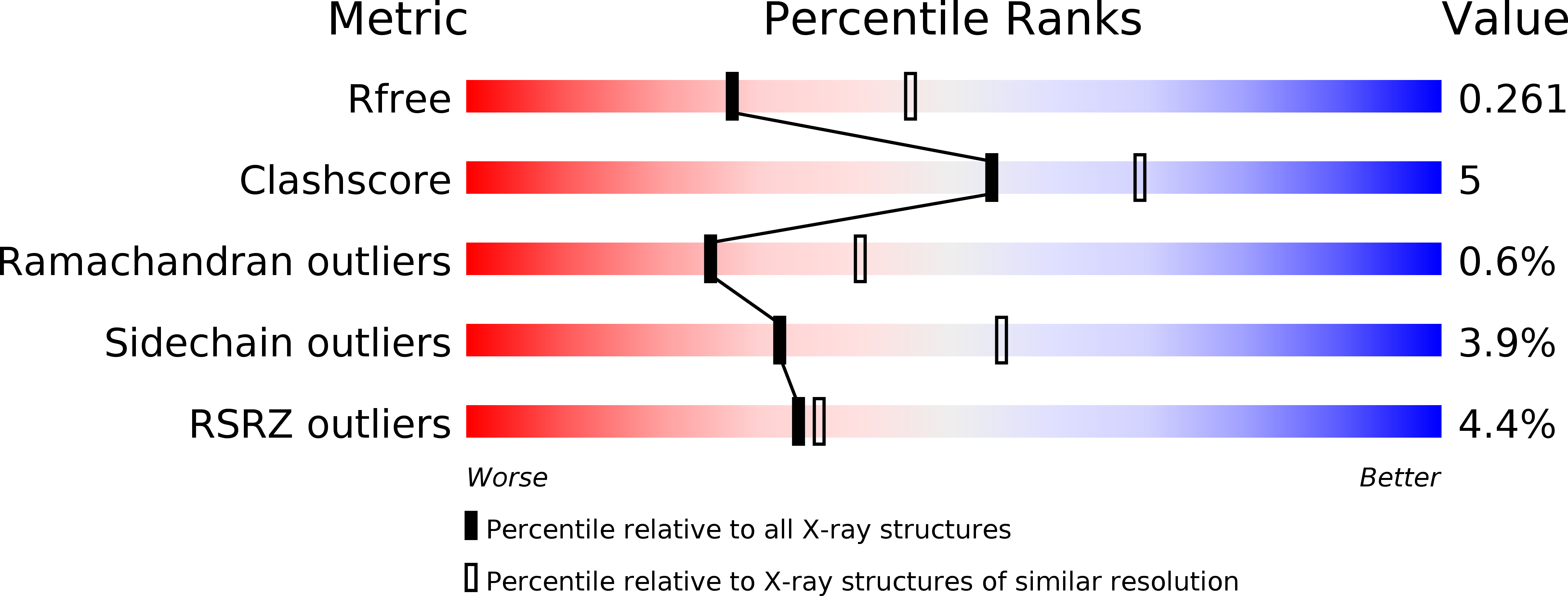
Resolution:
2.48 Å
R-Value Free:
0.25
R-Value Work:
0.19
R-Value Observed:
0.19
Space Group:
P 21 21 2
Macromolecular Entities
Polymer Type:polypeptide(L)
Molecule:FabOX117 light chain
Chain IDs:A, C
Chain Length:214
Number of Molecules:2
Biological Source:Homo sapiens
Polymer Type:polypeptide(L)
Molecule:FabOX117 heavy chain
Chain IDs:B, D
Chain Length:229
Number of Molecules:2
Biological Source:Homo sapiens
Structures with similar UniProt ID
Protein Blast
Polymer Type:polypeptide(L)
Molecule:Signal-regulatory protein gamma
Gene (Uniprot):SIRPG
Chain IDs:E, F
Chain Length:328
Number of Molecules:2
Biological Source:Homo sapiens
Modified Residue
Compound ID Chain ID Parent Comp ID Details 2D Image
ASN F ASN GLYCOSYLATION SITE
Primary Citation
Crystal structure of signal regulatory protein gamma (SIRP gamma) in complex with an antibody Fab fragment.
Bmc Struct.Biol. 13 13 13 (2013)
PMID: 23826770 DOI: 10.1186/1472-6807-13-13

Abstact

BACKGROUND Signal Regulatory Protein γ (SIRPγ) is a member of a closely related family of three cell surface receptors implicated in modulating immune/inflammatory responses. SIRPγ is expressed on T lymphocytes where it appears to be involved in the integrin-independent adhesion of lymphocytes to antigen-presenting cells. Here we describe the first full length structure of the extracellular region of human SIRPγ. RESULTS We obtained crystals of SIRPγ by making a complex of the protein with the Fab fragment of the anti-SIRP antibody, OX117, which also binds to SIRPα and SIRPβ. We show that the epitope for FabOX117 is formed at the interface of the first and second domains of SIRPγ and comprises residues which are conserved between all three SIRPs. The FabOX117 binding site is distinct from the region in domain 1 which interacts with CD47, the physiological ligand for both SIRPγ and SIRPα but not SIRPβ. Comparison of the three domain structures of SIRPγ and SIRPα showed that these receptors can adopt different overall conformations due to the flexibility of the linker between the first two domains. SIRPγ in complex with FabOX117 forms a dimer in the crystal. Binding to the Fab fixes the position of domain 1 relative to domains 2/3 exposing a surface which favours formation of a homotypic dimer. However, the interaction appears to be relatively weak since only monomers of SIRPγ were observed in sedimentation velocity analytical ultracentrifugation of the protein alone. Studies of complex formation by equilibrium ultracentrifugation showed that only a 1:1 complex of SIRPγ: FabOX117 was formed with a dissociation constant in the low micromolar range (Kd = 1.2 +/- 0.3 μM). CONCLUSION The three-domain extracellular regions of SIRPs are structurally conserved but show conformational flexibility in the disposition of the amino terminal ligand-binding Ig domain relative to the two membrane proximal Ig domains. Binding of a cross-reactive anti-SIRP Fab fragment to SIRPγ stabilises a conformation that favours SIRP dimer formation in the crystal structure, though this interaction does not appear sufficiently stable to be observed in solution.

Legend

Protein

Chemical

Disease

Primary Citation of related structures