4I2R image
Entry Detail
PDB ID:
4I2R
Keywords:
Title:
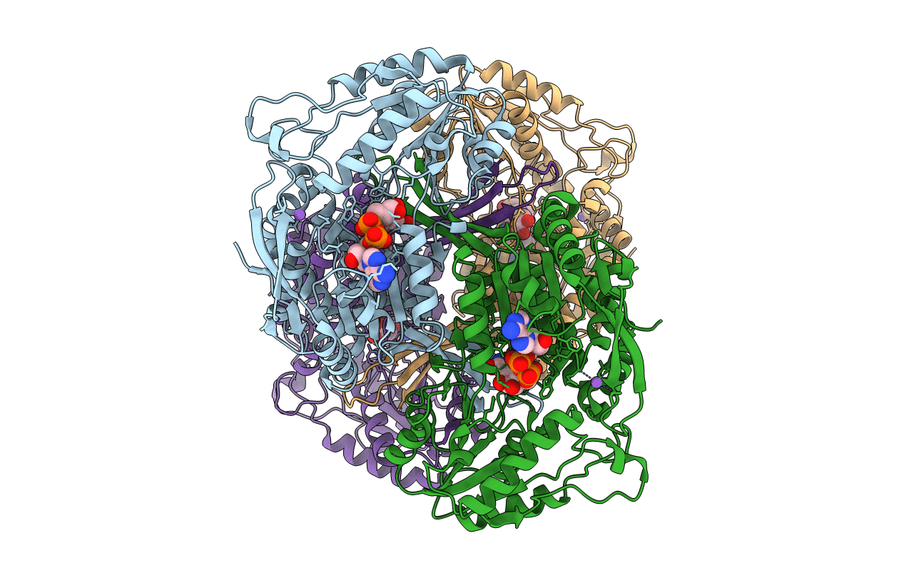
2.15 Angstroms X-ray crystal structure of NAD- and alternative substrate-bound 2-aminomuconate 6-semialdehyde dehydrogenase from Pseudomonas fluorescens
Biological Source:
Source Organism:
Host Organism:
PDB Version:
Deposition Date:
2012-11-22
Release Date:
2014-05-21
Method Details:
Experimental Method:
Resolution:
2.15 Å
R-Value Free:
0.23
R-Value Work:
0.17
R-Value Observed:
0.18
Space Group:
P 21 21 21
Macromolecular Entities
Polymer Type:polypeptide(L)
Description:2-aminomuconate 6-semialdehyde dehydrogenase
Chain IDs:A, B, C, D
Chain Length:500
Number of Molecules:4
Biological Source:Pseudomonas fluorescens
Primary Citation
Crystallographic and spectroscopic snapshots reveal a dehydrogenase in action.
Nat Commun 6 5935 5935 (2015)
PMID: 25565451 DOI: 10.1038/ncomms6935

Abstact

Aldehydes are ubiquitous intermediates in metabolic pathways and their innate reactivity can often make them quite unstable. There are several aldehydic intermediates in the metabolic pathway for tryptophan degradation that can decay into neuroactive compounds that have been associated with numerous neurological diseases. An enzyme of this pathway, 2-aminomuconate-6-semialdehyde dehydrogenase, is responsible for 'disarming' the final aldehydic intermediate. Here we show the crystal structures of a bacterial analogue enzyme in five catalytically relevant forms: resting state, one binary and two ternary complexes, and a covalent, thioacyl intermediate. We also report the crystal structures of a tetrahedral, thiohemiacetal intermediate, a thioacyl intermediate and an NAD(+)-bound complex from an active site mutant. These covalent intermediates are characterized by single-crystal and solution-state electronic absorption spectroscopy. The crystal structures reveal that the substrate undergoes an E/Z isomerization at the enzyme active site before an sp(3)-to-sp(2) transition during enzyme-mediated oxidation.

Legend

Protein

Chemical

Disease

Primary Citation of related structures