Deposition Date
2012-11-21
Release Date
2013-01-30
Last Version Date
2024-11-06
Entry Detail
PDB ID:
4I1S
Keywords:
Title:
Melanoma differentiation associated protein-5 Helicase domain complex with inhibitor Non-structural protein V
Biological Source:
Source Organism(s):
Sus scrofa (Taxon ID: 9823)
Simian virus 5 (Taxon ID: 11208)
Simian virus 5 (Taxon ID: 11208)
Expression System(s):
Method Details:
Experimental Method:
Resolution:
2.29 Å
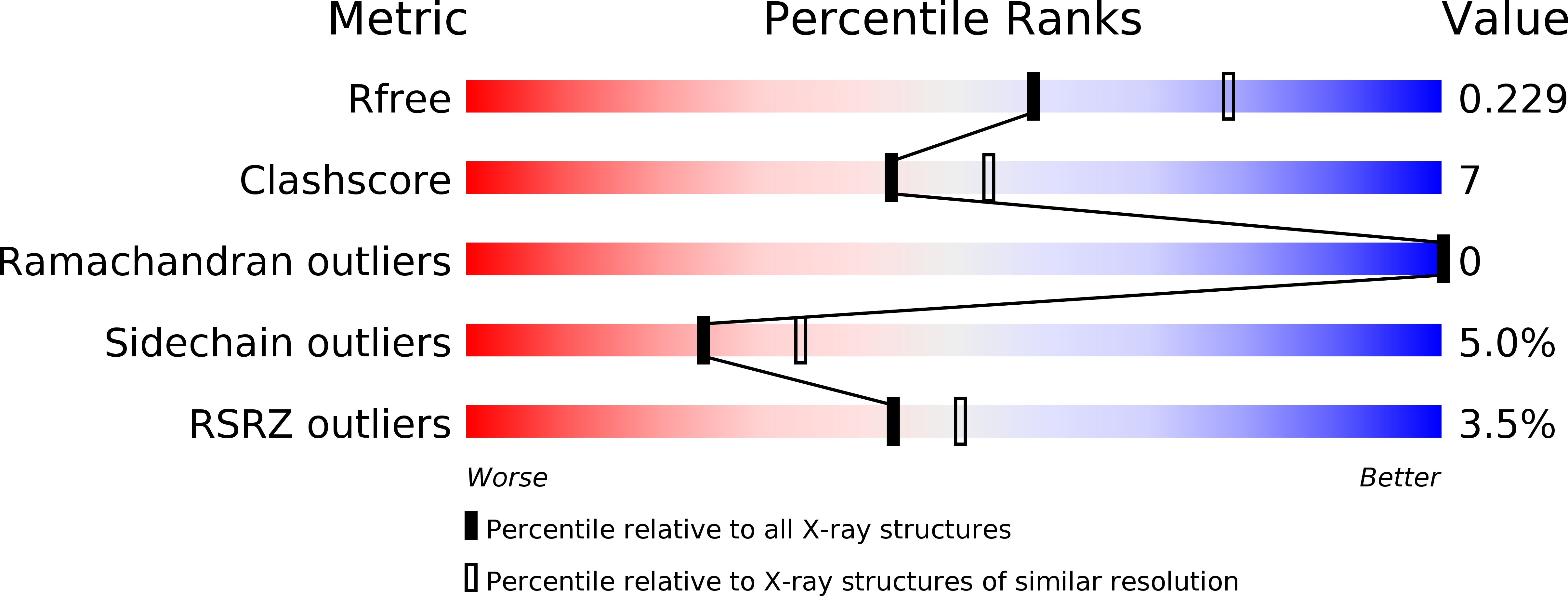
R-Value Free:
0.22
R-Value Work:
0.17
R-Value Observed:
0.18
Space Group:
P 21 21 21