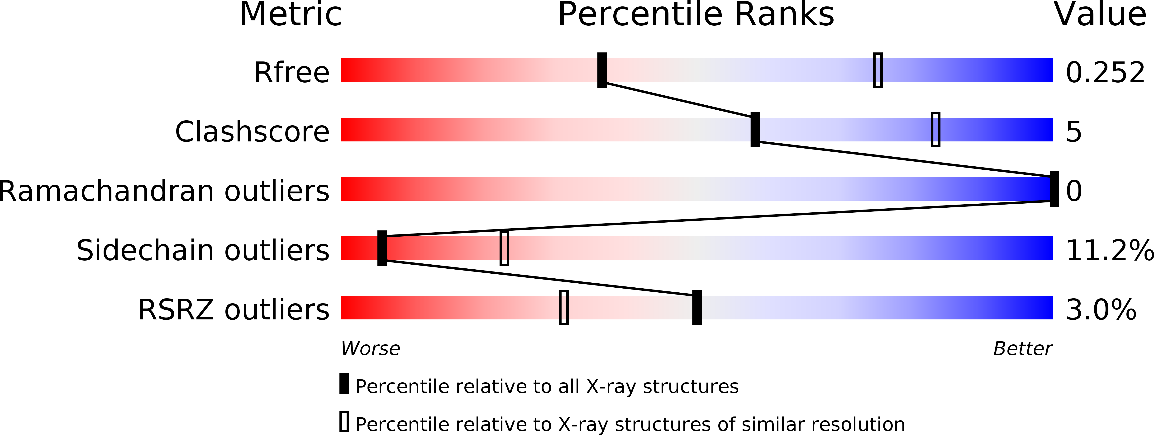
4I0K image
Deposition Date 2012-11-16
Release Date 2012-12-05
Last Version Date 2024-10-16
Entry Detail
PDB ID:
4I0K
Keywords:
Title:
Crystal structure of murine B7-H3 extracellular domain
Biological Source:
Source Organism:
Mus musculus (Taxon ID: 10090)
Method Details:
Experimental Method:
Resolution:
2.97 Å
R-Value Free:
0.24
R-Value Work:
0.21
R-Value Observed:
0.21
Space Group:
P 61 2 2
Macromolecular Entities
Polymer Type:polypeptide(L)
Molecule:CD276 antigen
Gene (Uniprot):Cd276
Chain IDs:A
Chain Length:222
Number of Molecules:1
Biological Source:Mus musculus
Modified Residue
Compound ID Chain ID Parent Comp ID Details 2D Image
ASN A ASN GLYCOSYLATION SITE
Primary Citation
Structure and T cell inhibition properties of B7 family member, B7-H3.
Structure 21 707 717 (2013)
PMID: 23583036 DOI: 10.1016/j.str.2013.03.003

Abstact

T cell activity is controlled by a combination of antigen-dependent signaling through the T cell receptor and a set of auxiliary signals delivered through antigen-independent interactions, including the recognition of the B7 family of ligands. B7-H3 is a recently identified B7 family member that is strongly overexpressed in a range of cancers and correlates with poor prognosis. We report the crystal structure of murine B7-H3 at a 3 Å resolution, which provides a model for the organization of the IgV and IgC domains within the ectodomain. We demonstrate that B7-H3 inhibits T cell proliferation and show that the FG loop of the IgV domain plays a critical role in this function. B7-H3 crystallized as an unusual dimer arising from the exchange of the G strands in the IgV domains of partner molecules. This arrangement, in combination with previous reports, highlights the dynamic nature and plasticity of the immunoglobulin fold.

Legend

Protein

Chemical

Disease

Primary Citation of related structures