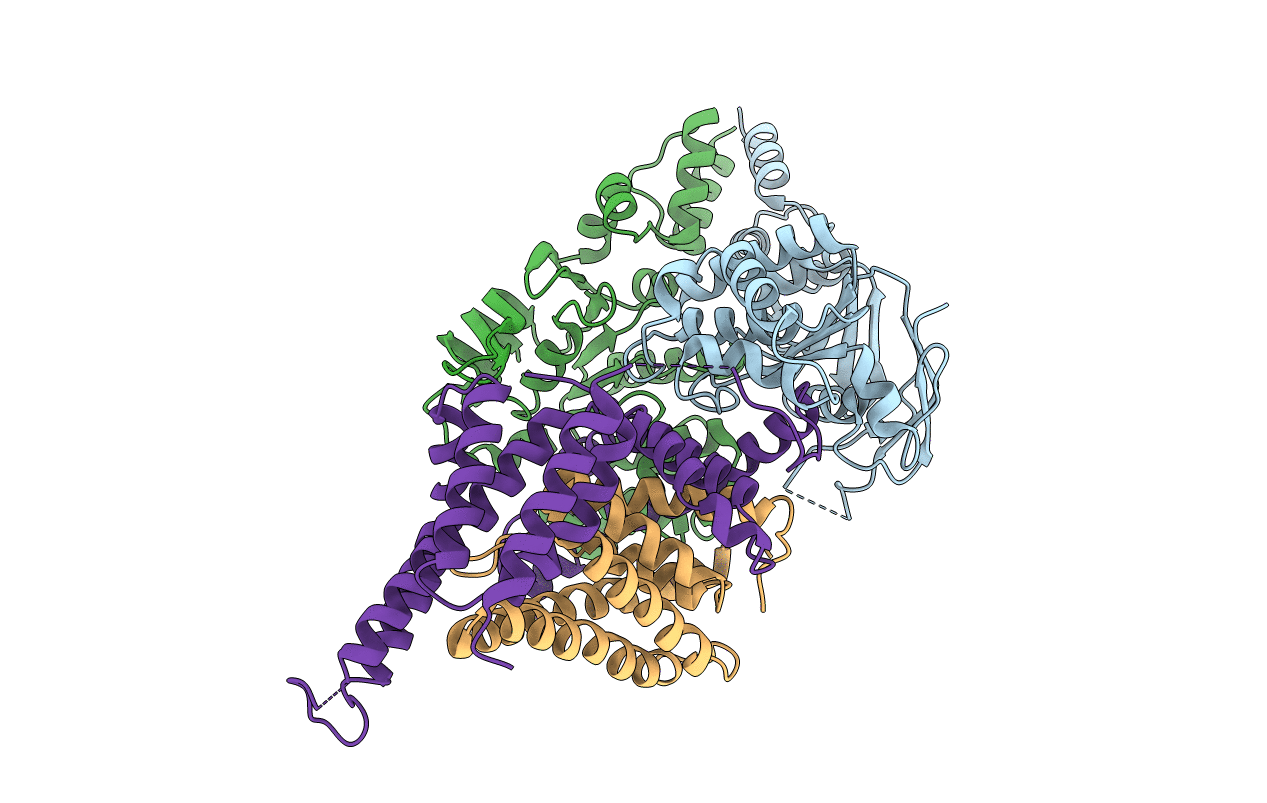
4HZU image
Entry Detail
PDB ID:
4HZU
Title:
Structure of a bacterial energy-coupling factor transporter
Biological Source:
Source Organism:
Host Organism:
PDB Version:
Deposition Date:
2012-11-15
Release Date:
2013-04-17
Method Details:
Experimental Method:
Resolution:
3.53 Å
R-Value Free:
0.29
R-Value Work:
0.24
R-Value Observed:
0.24
Space Group:
P 21 21 21
Macromolecular Entities
Polymer Type:polypeptide(L)
Description:Energy-coupling factor transporter ATP-binding protein EcfA 2
Chain IDs:B (auth: A)
Chain Length:279
Number of Molecules:1
Biological Source:Lactobacillus brevis
Polymer Type:polypeptide(L)
Description:Energy-coupling factor transporter ATP-binding protein EcfA 1
Chain IDs:A (auth: B)
Chain Length:290
Number of Molecules:1
Biological Source:Lactobacillus brevis
Polymer Type:polypeptide(L)
Description:Predicted membrane protein
Chain IDs:D (auth: S)
Chain Length:166
Number of Molecules:1
Biological Source:Lactobacillus brevis
Polymer Type:polypeptide(L)
Description:Energy-coupling factor transporter transmembrane protein EcfT
Chain IDs:C (auth: T)
Chain Length:266
Number of Molecules:1
Biological Source:Lactobacillus brevis
Ligand Molecules
Primary Citation
Structure of a bacterial energy-coupling factor transporter.
Nature 497 272 276 (2013)
PMID: 23584587 DOI: 10.1038/nature12045

Abstact

The energy-coupling factor (ECF) transporters constitute a novel family of conserved membrane transporters in prokaryotes that have a similar domain organization to the ATP-binding cassette transporters. Each ECF transporter comprises a pair of cytosolic ATPases (the A and A' components, or EcfA and EcfA'), a membrane-embedded substrate-binding protein (the S component, or EcfS) and a transmembrane energy-coupling component (the T component, or EcfT) that links the EcfA-EcfA' subcomplex to EcfS. The structure and transport mechanism of the quaternary ECF transporter remain largely unknown. Here we report the crystal structure of a nucleotide-free ECF transporter from Lactobacillus brevis at a resolution of 3.5 Å. The T component has a horseshoe-shaped open architecture, with five α-helices as transmembrane segments and two cytoplasmic α-helices as coupling modules connecting to the A and A' components. Strikingly, the S component, thought to be specific for hydroxymethyl pyrimidine, lies horizontally along the lipid membrane and is bound exclusively by the five transmembrane segments and the two cytoplasmic helices of the T component. These structural features suggest a plausible working model for the transport cycle of the ECF transporters.

Legend

Protein

Chemical

Disease

Primary Citation of related structures