
Deposition Date
2012-11-14
Release Date
2012-12-12
Last Version Date
2024-10-16
Entry Detail
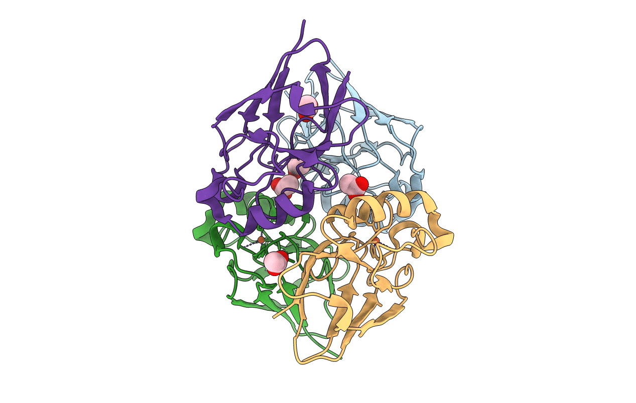
PDB ID:
4HZ1
Keywords:
Title:
Crystal Structure of Pseudomonas aeruginosa azurin with iron(II) at the copper-binding site.
Biological Source:
Source Organism(s):
Pseudomonas aeruginosa (Taxon ID: 208964)
Expression System(s):
Method Details:
Experimental Method:
Resolution:
2.20 Å
R-Value Free:
0.27
R-Value Work:
0.24
Space Group:
P 21 21 21
