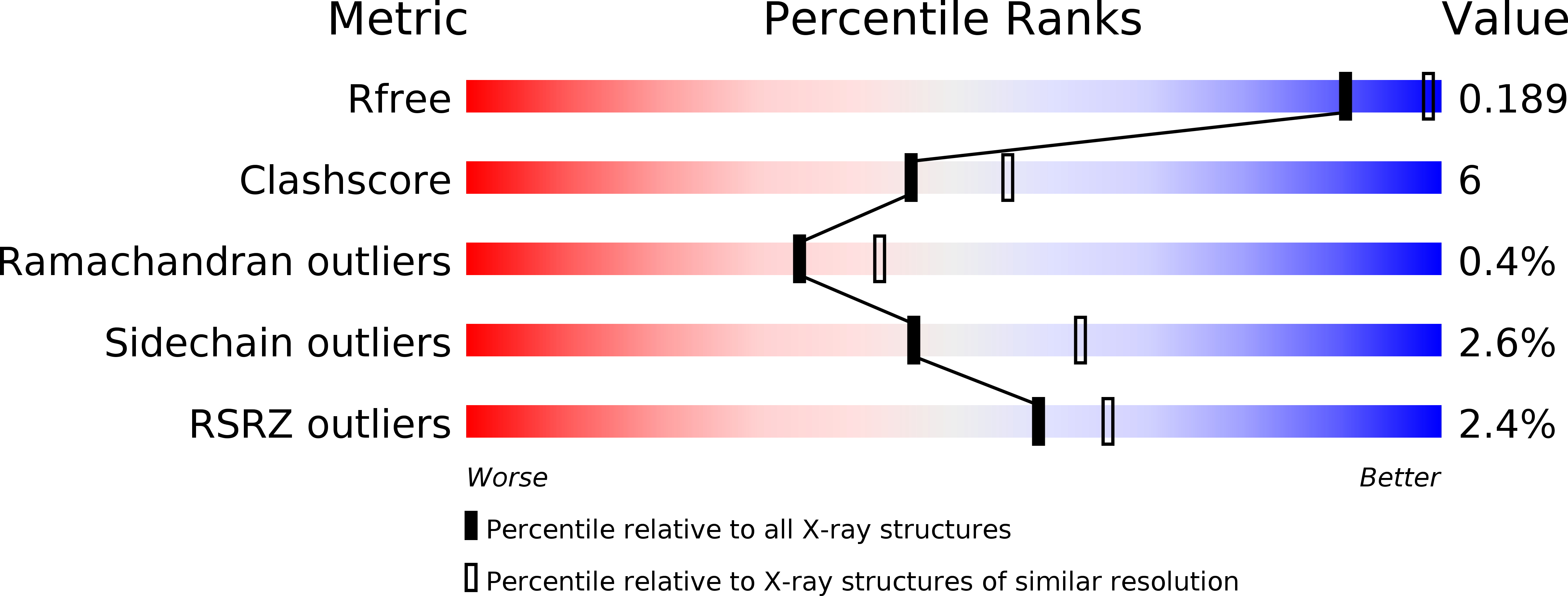
4HYV image
Deposition Date 2012-11-14
Release Date 2013-09-11
Last Version Date 2024-02-28
Entry Detail
PDB ID:
4HYV
Keywords:
Title:
Pyruvate kinase (PYK) from Trypanosoma brucei in the presence of Magnesium, PEP and F26BP
Biological Source:
Source Organism:
Host Organism:
Method Details:
Experimental Method:
Resolution:
2.30 Å
R-Value Free:
0.18
R-Value Work:
0.14
R-Value Observed:
0.14
Space Group:
I 2 2 2
Macromolecular Entities
Polymer Type:polypeptide(L)
Molecule:Pyruvate kinase 1
Gene (Uniprot):PYK1
Chain IDs:A, B
Chain Length:499
Number of Molecules:2
Biological Source:Trypanosoma brucei brucei
Primary Citation
`In crystallo' substrate binding triggers major domain movements and reveals magnesium as a co-activator of Trypanosoma brucei pyruvate kinase.
Acta Crystallogr.,Sect.D 69 1768 1779 (2013)
PMID: 23999300 DOI: 10.1107/S0907444913013875

Abstact

The active site of pyruvate kinase (PYK) is located between the AC core of the enzyme and a mobile lid corresponding to domain B. Many PYK structures have already been determined, but the first `effector-only' structure and the first with PEP (the true natural substrate) are now reported for the enzyme from Trypanosoma brucei. PEP soaked into crystals of the enzyme with bound allosteric activator fructose 2,6-bisphosphate (F26BP) and Mg(2+) triggers a substantial 23° rotation of the B domain `in crystallo', resulting in a partially closed active site. The interplay of side chains with Mg(2+) and PEP may explain the mechanism of the domain movement. Furthermore, it is apparent that when F26BP is present but PEP is absent Mg(2+) occupies a position that is distinct from the two canonical Mg(2+)-binding sites at the active site. This third site is adjacent to the active site and involves the same amino-acid side chains as in canonical site 1 but in altered orientations. Site 3 acts to sequester Mg(2+) in a `priming' position such that the enzyme is maintained in its R-state conformation. In this way, Mg(2+) cooperates with F26BP to ensure that the enzyme is in a conformation that has a high affinity for the substrate.

Legend

Protein

Chemical

Disease

Primary Citation of related structures