
Deposition Date
2012-11-14
Release Date
2013-06-26
Last Version Date
2023-09-20
Entry Detail
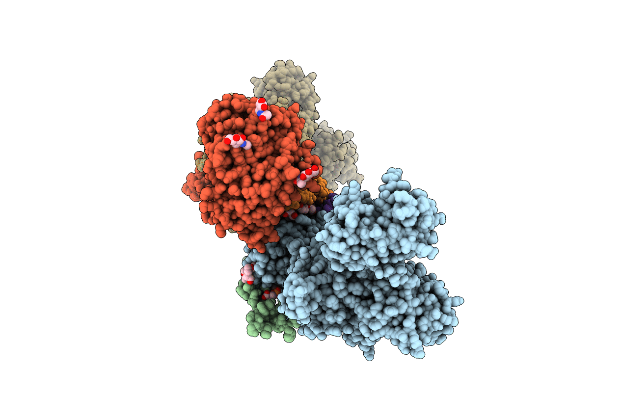
PDB ID:
4HYT
Keywords:
Title:
Na,K-ATPase in the E2P state with bound ouabain and Mg2+ in the cation-binding site
Biological Source:
Source Organism(s):
Sus scrofa (Taxon ID: 9823)
Method Details:
Experimental Method:
Resolution:
3.40 Å
R-Value Free:
0.24
R-Value Work:
0.22
R-Value Observed:
0.22
Space Group:
P 21 21 21
