
Deposition Date
2012-11-14
Release Date
2013-05-29
Last Version Date
2024-11-06
Entry Detail
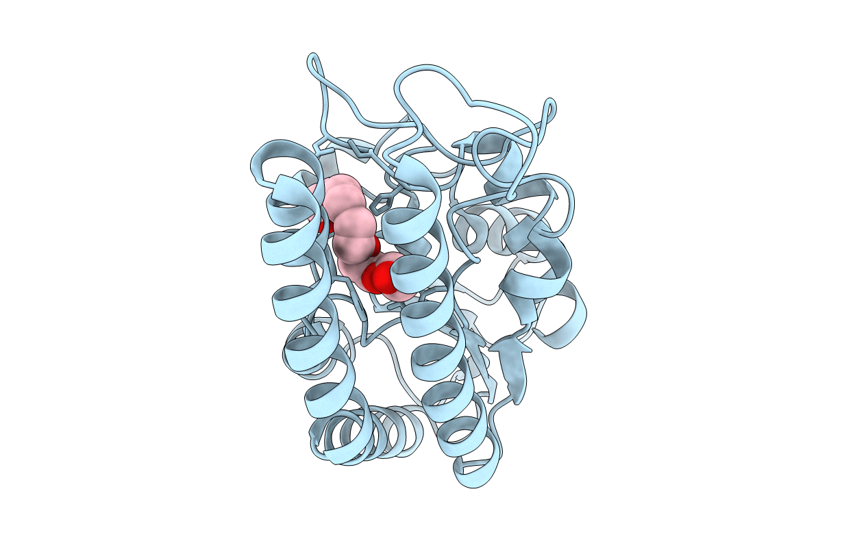
PDB ID:
4HYQ
Keywords:
Title:
Crystal structure of phospholipase A1 from Streptomyces albidoflavus NA297
Biological Source:
Source Organism:
Streptomyces albidoflavus (Taxon ID: 1886)
Host Organism:
Method Details:
Experimental Method:
Resolution:
1.75 Å
R-Value Free:
0.22
R-Value Work:
0.18
R-Value Observed:
0.18
Space Group:
P 31 2 1
