Deposition Date
2012-11-13
Release Date
2012-12-19
Last Version Date
2024-03-20
Entry Detail
PDB ID:
4HYD
Keywords:
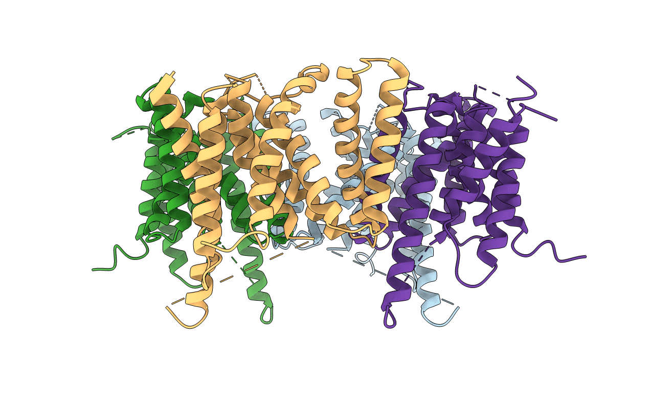
Title:
Structure of a presenilin family intramembrane aspartate protease in C2221 space group
Biological Source:
Source Organism(s):
Methanoculleus marisnigri JR1 (Taxon ID: 368407)
Expression System(s):
Method Details:
Experimental Method:
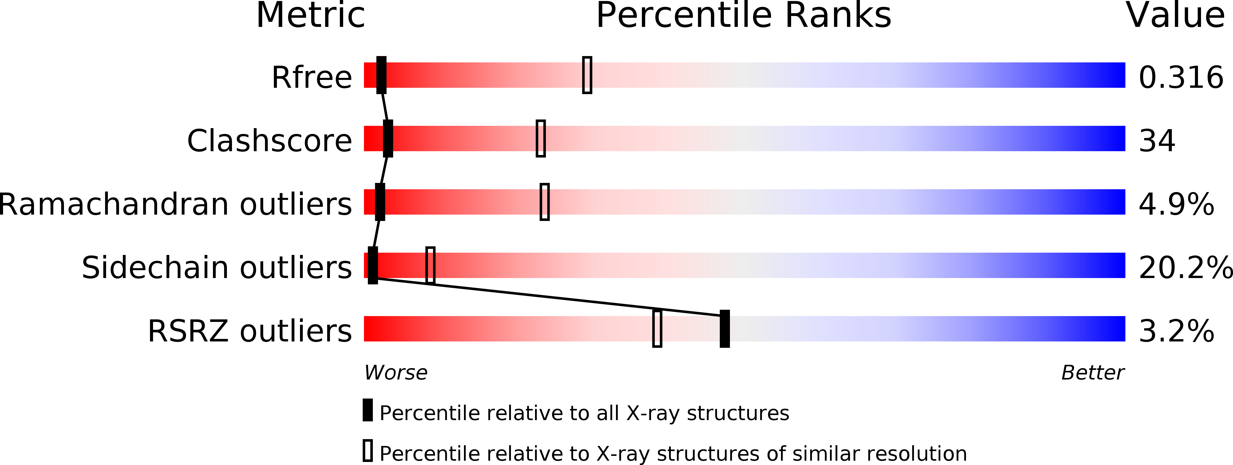
Resolution:
3.80 Å
R-Value Free:
0.30
R-Value Work:
0.30
R-Value Observed:
0.30
Space Group:
C 2 2 21