4HUT image
Deposition Date 2012-11-04
Release Date 2013-01-23
Last Version Date 2023-09-20
Entry Detail
PDB ID:
4HUT
Keywords:
Title:
Structure of ATP:co(I)rrinoid adenosyltransferase (CobA) from Salmonella enterica in complex with four and five-coordinate cob(II)alamin and ATP
Biological Source:
Method Details:
Experimental Method:
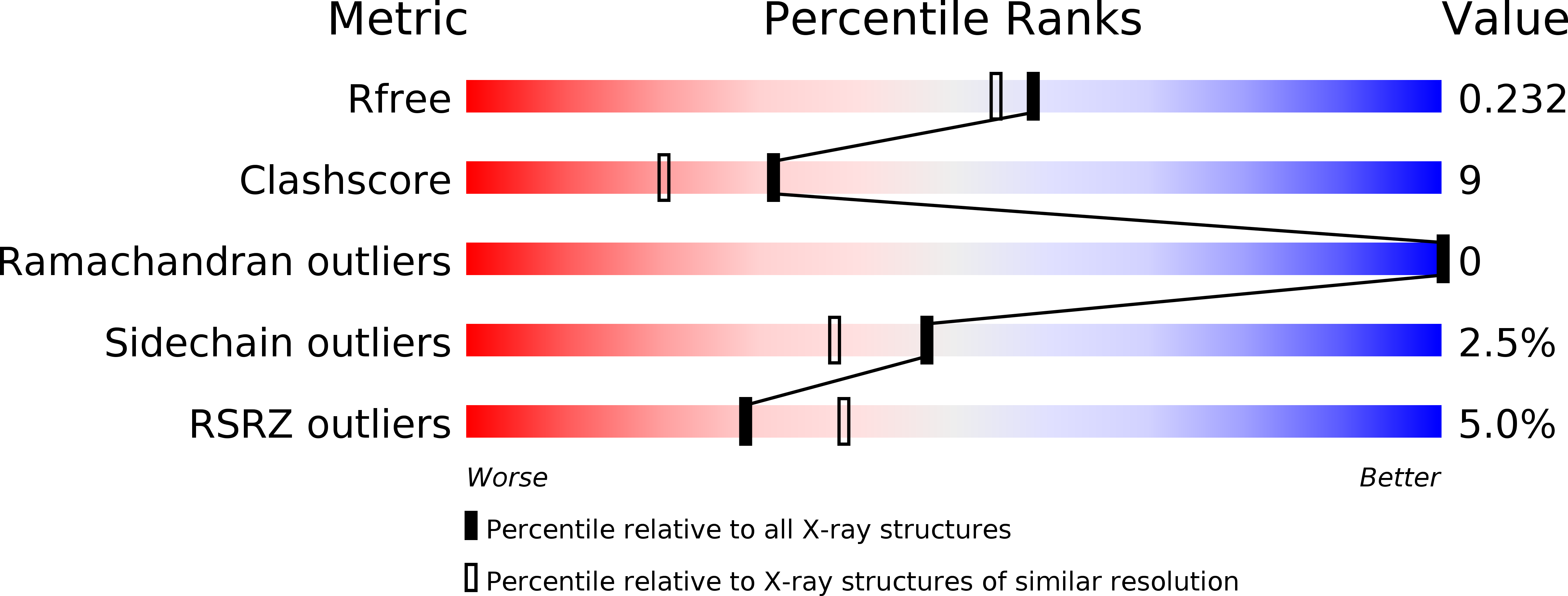
Resolution:
1.95 Å
R-Value Free:
0.23
R-Value Work:
0.17
R-Value Observed:
0.17
Space Group:
P 21 21 21
Macromolecular Entities
Polymer Type:polypeptide(L)
Molecule:Cob(I)yrinic acid a,c-diamide adenosyltransferase
Gene (Uniprot):btuR
Chain IDs:A, B
Chain Length:191
Number of Molecules:2
Biological Source:Salmonella enterica subsp. enterica serovar Typhimurium
Primary Citation
Structural Insights into the Mechanism of Four-Coordinate Cob(II)alamin Formation in the Active Site of the Salmonella enterica ATP:Co(I)rrinoid Adenosyltransferase Enzyme: Critical Role of Residues Phe91 and Trp93.
Biochemistry 51 9647 9657 (2012)
PMID: 23148601 DOI: 10.1021/bi301378d

Abstact

ATP:co(I)rrinoid adenosyltransferases (ACATs) are enzymes that catalyze the formation of adenosylcobalamin (AdoCbl, coenzyme B(12)) from cobalamin and ATP. There are three families of ACATs, namely, CobA, EutT, and PduO. In Salmonella enterica, CobA is the housekeeping enzyme that is required for de novo AdoCbl synthesis and for salvaging incomplete precursors and cobalamin from the environment. Here, we report the crystal structure of CobA in complex with ATP, four-coordinate cobalamin, and five-coordinate cobalamin. This provides the first crystallographic evidence of the existence of cob(II)alamin in the active site of CobA. The structure suggests a mechanism in which the enzyme adopts a closed conformation and two residues, Phe91 and Trp93, displace 5,6-dimethylbenzimidazole, the lower nucleotide ligand base of cobalamin, to generate a transient four-coordinate cobalamin, which is critical in the formation of the AdoCbl Co-C bond. In vivo and in vitro mutational analyses of Phe91 and Trp93 emphasize the important role of bulky hydrophobic side chains in the active site. The proposed manner in which CobA increases the redox potential of the cob(II)alamin/cob(I)alamin couple to facilitate formation of the Co-C bond appears to be analogous to that utilized by the PduO-type ACATs, where in both cases the polar coordination of the lower ligand to the cobalt ion is eliminated by placing that face of the corrin ring adjacent to a cluster of bulky hydrophobic side chains.

Legend

Protein

Chemical

Disease

Primary Citation of related structures