
Deposition Date
2012-11-02
Release Date
2013-08-28
Last Version Date
2023-09-20
Entry Detail
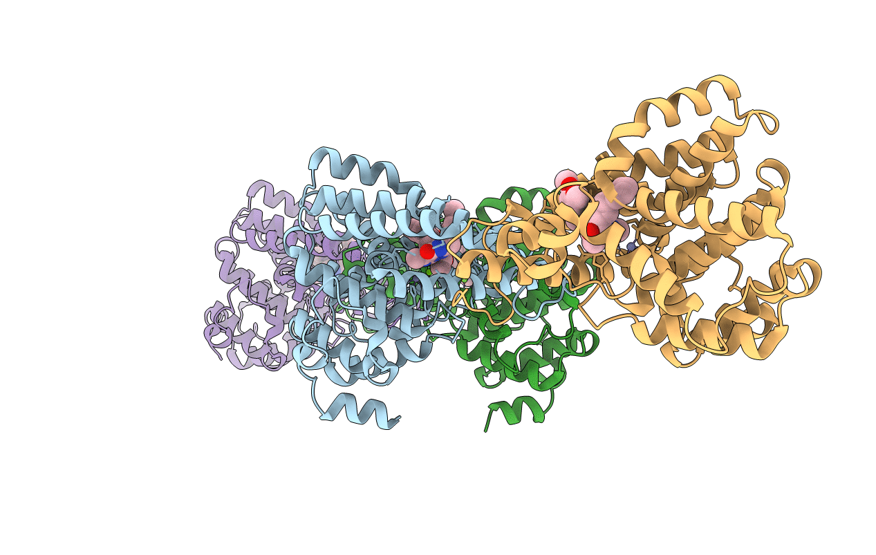
PDB ID:
4HTX
Keywords:
Title:
Crystal structure of PDE2 catalytic domain in complex with BAY60-7550
Biological Source:
Source Organism(s):
Homo sapiens (Taxon ID: 9606)
Expression System(s):
Method Details:
Experimental Method:
Resolution:
1.90 Å
R-Value Free:
0.22
R-Value Work:
0.17
R-Value Observed:
0.17
Space Group:
P 1
