Deposition Date
2012-11-01
Release Date
2013-05-01
Last Version Date
2024-02-28
Entry Detail
PDB ID:
4HTS
Keywords:
Title:
Crystal Structure of Twin Arginine Translocase Receptor- TatC
Biological Source:
Source Organism(s):
Aquifex aeolicus VF5 (Taxon ID: 224324)
Expression System(s):
Method Details:
Experimental Method:
Resolution:
4.00 Å
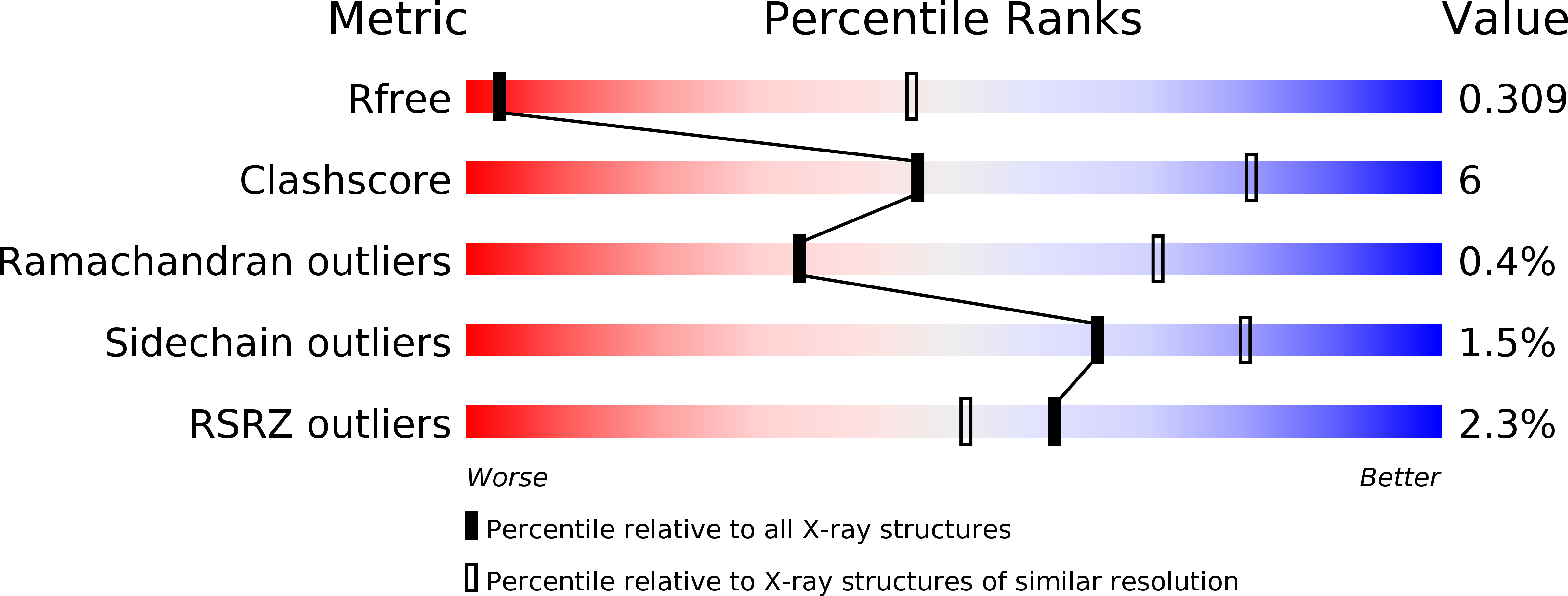
R-Value Free:
0.32
R-Value Work:
0.28
R-Value Observed:
0.29
Space Group:
P 41 2 2