Deposition Date
2012-11-01
Release Date
2012-11-28
Last Version Date
2023-09-20
Entry Detail
PDB ID:
4HTJ
Keywords:
Title:
Crystallographic structure of the membrane-proximal ectodomain of the human receptor-type protein-tyrosine phosphatase phogrin at pH 4.6
Biological Source:
Source Organism(s):
Homo sapiens (Taxon ID: 9606)
Expression System(s):
Method Details:
Experimental Method:
Resolution:
2.01 Å
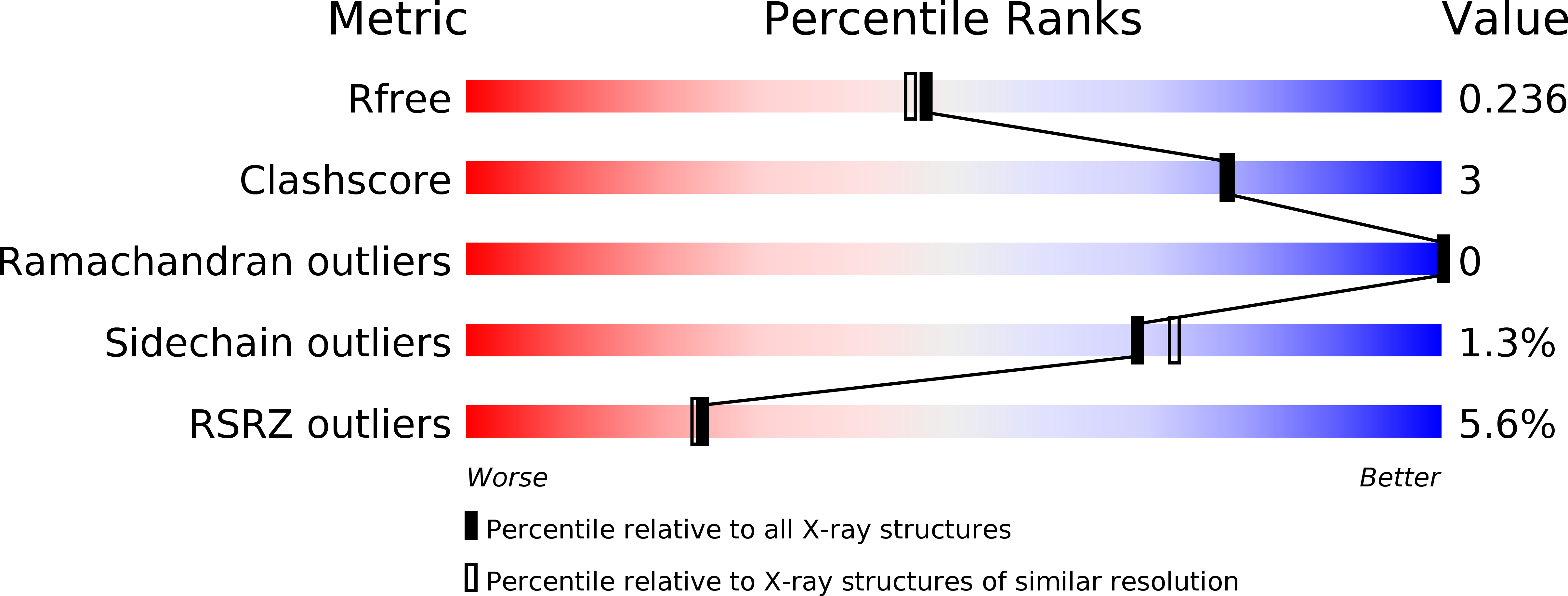
R-Value Free:
0.23
R-Value Work:
0.20
R-Value Observed:
0.20
Space Group:
P 61 2 2