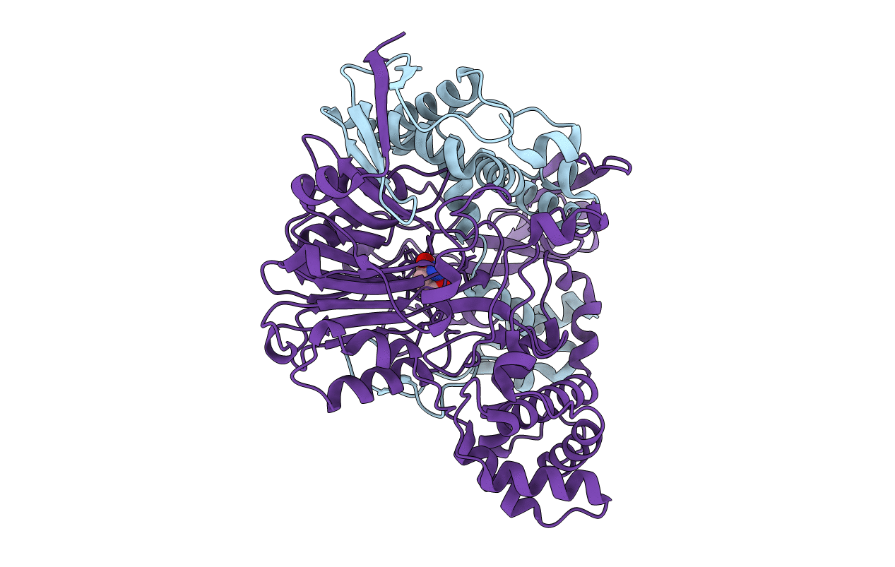
4HST image
Entry Detail
PDB ID:
4HST
Keywords:
Title:
Crystal structure of a double mutant of a class III engineered cephalosporin acylase
Biological Source:
Source Organism:
Host Organism:
PDB Version:
Deposition Date:
2012-10-30
Release Date:
2013-02-27
Method Details:
Experimental Method:
Resolution:
1.57 Å
R-Value Free:
0.16
R-Value Work:
0.11
R-Value Observed:
0.12
Space Group:
P 21 21 21
Macromolecular Entities
Polymer Type:polypeptide(L)
Description:glutaryl-7-aminocephalosporanic acid acylase alpha chain
Chain IDs:A
Chain Length:229
Number of Molecules:1
Biological Source:Pseudomonas
Polymer Type:polypeptide(L)
Description:glutaryl-7-aminocephalosporanic acid acylase beta chain
Chain IDs:B
Chain Length:543
Number of Molecules:1
Biological Source:Pseudomonas
Ligand Molecules
Primary Citation
Structure of a class III engineered cephalosporin acylase: comparisons with class I acylase and implications for differences in substrate specificity and catalytic activity.
Biochem.J. 451 217 226 (2013)
PMID: 23373797 DOI: 10.1042/BJ20121715

Abstact

The crystal structure of the wild-type form of glutaryl-7-ACA (7-aminocephalosporanic acid) acylase from Pseudomonas N176 and a double mutant of the protein (H57βS/H70βS) that displays enhanced catalytic efficiency on cephalosporin C over glutaryl-7-aminocephalosporanic acid has been determined. The structures show a heterodimer made up of an α-chain (229 residues) and a β-chain (543 residues) with a deep cavity, which constitutes the active site. Comparison of the wild-type and mutant structures provides insights into the molecular reasons for the observed enhanced specificity on cephalosporin C over glutaryl-7-aminocephalosporanic acid and offers the basis to evolve a further improved enzyme variant. The nucleophilic catalytic serine residue, Ser(1β), is situated at the base of the active site cavity. The electron density reveals a ligand covalently bound to the catalytic serine residue, such that a tetrahedral adduct is formed. This is proposed to mimic the transition state of the enzyme for both the maturation step and the catalysis of the substrates. A view of the transition state configuration of the enzyme provides important insights into the mechanism of substrate binding and catalysis.

Legend

Protein

Chemical

Disease

Primary Citation of related structures