4HSR image
Deposition Date 2012-10-30
Release Date 2013-02-27
Last Version Date 2024-02-28
Entry Detail
PDB ID:
4HSR
Keywords:
Title:
Crystal Structure of a class III engineered cephalosporin acylase
Biological Source:
Source Organism:
Pseudomonas (Taxon ID: 286)
Host Organism:
Method Details:
Experimental Method:
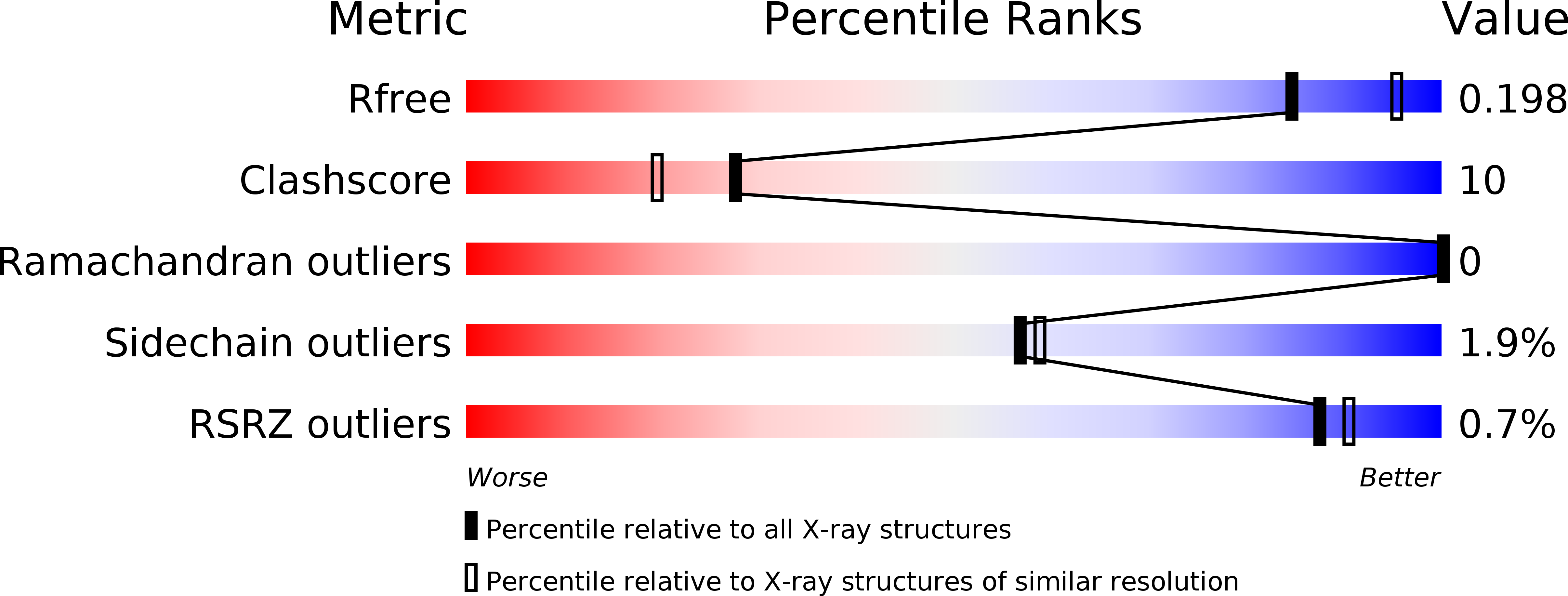
Resolution:
2.13 Å
R-Value Free:
0.19
R-Value Work:
0.15
R-Value Observed:
0.15
Space Group:
P 21 21 21
Macromolecular Entities
Polymer Type:polypeptide(L)
Molecule:glutaryl-7-aminocephalosporanic acid acylase alpha chain
Chain IDs:A
Chain Length:229
Number of Molecules:1
Biological Source:Pseudomonas
Polymer Type:polypeptide(L)
Molecule:glutaryl-7-aminocephalosporanic acid acylase beta chain
Chain IDs:B
Chain Length:543
Number of Molecules:1
Biological Source:Pseudomonas
Ligand Molecules
Primary Citation
Structure of a class III engineered cephalosporin acylase: comparisons with class I acylase and implications for differences in substrate specificity and catalytic activity.
Biochem.J. 451 217 226 (2013)
PMID: 23373797 DOI: 10.1042/BJ20121715

Abstact

The crystal structure of the wild-type form of glutaryl-7-ACA (7-aminocephalosporanic acid) acylase from Pseudomonas N176 and a double mutant of the protein (H57βS/H70βS) that displays enhanced catalytic efficiency on cephalosporin C over glutaryl-7-aminocephalosporanic acid has been determined. The structures show a heterodimer made up of an α-chain (229 residues) and a β-chain (543 residues) with a deep cavity, which constitutes the active site. Comparison of the wild-type and mutant structures provides insights into the molecular reasons for the observed enhanced specificity on cephalosporin C over glutaryl-7-aminocephalosporanic acid and offers the basis to evolve a further improved enzyme variant. The nucleophilic catalytic serine residue, Ser(1β), is situated at the base of the active site cavity. The electron density reveals a ligand covalently bound to the catalytic serine residue, such that a tetrahedral adduct is formed. This is proposed to mimic the transition state of the enzyme for both the maturation step and the catalysis of the substrates. A view of the transition state configuration of the enzyme provides important insights into the mechanism of substrate binding and catalysis.

Legend

Protein

Chemical

Disease

Primary Citation of related structures
Feedback Form
Name
Email
Institute
Feedback