4HPU image
Deposition Date 2012-10-24
Release Date 2013-03-20
Last Version Date 2024-11-06
Entry Detail
PDB ID:
4HPU
Title:
Crystal structure of the catalytic subunit of cAMP-dependent protein kinase displaying partial phosphoryl transfer of AMP-PNP onto a substrate peptide
Biological Source:
Source Organism:
Mus musculus (Taxon ID: 10090)
Host Organism:
Method Details:
Experimental Method:
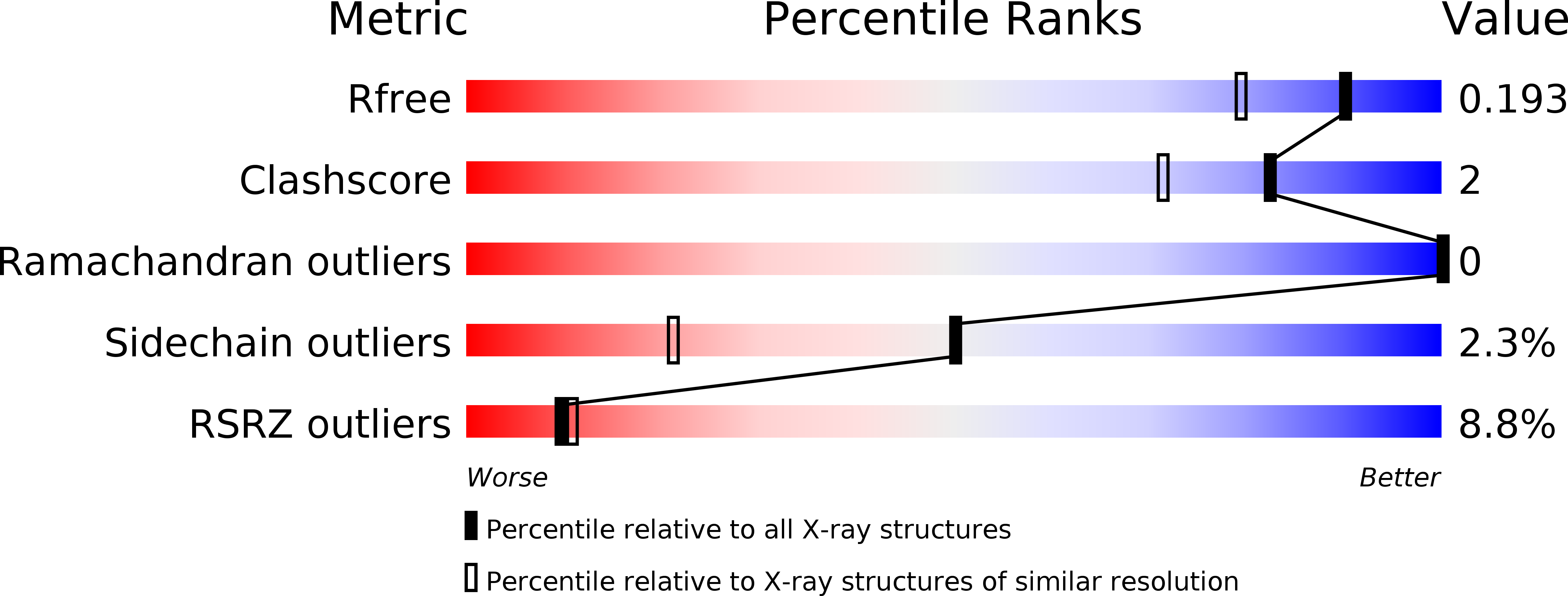
Resolution:
1.55 Å
R-Value Free:
0.19
R-Value Work:
0.17
R-Value Observed:
0.17
Space Group:
P 21 21 21
Macromolecular Entities
Structures with similar UniProt ID
Protein Blast
Polymer Type:polypeptide(L)
Molecule:cAMP-dependent protein kinase catalytic subunit alpha
Gene (Uniprot):Prkaca
Chain IDs:A (auth: E)
Chain Length:350
Number of Molecules:1
Biological Source:Mus musculus
Structures with similar UniProt ID
Protein Blast
Polymer Type:polypeptide(L)
Molecule:cAMP-dependent protein kinase inhibitor alpha
Gene (Uniprot):Pkia
Mutagens:N20A, A21S
Chain IDs:B (auth: I)
Chain Length:20
Number of Molecules:1
Biological Source:Mus musculus
Modified Residue
Compound ID Chain ID Parent Comp ID Details 2D Image
SEP A SER PHOSPHOSERINE
TPO A THR PHOSPHOTHREONINE
Primary Citation
Phosphoryl transfer by protein kinase a is captured in a crystal lattice.
J.Am.Chem.Soc. 135 4788 4798 (2013)
PMID: 23458248 DOI: 10.1021/ja312237q

Abstact

The catalytic (C) subunit of cAMP-dependent protein kinase (PKA) is a serine/threonine kinase responsible for most of the effects of cAMP signaling, and PKA serves as a prototype for the entire kinase family. Despite multiple studies of PKA, the steps involved in phosphoryl transfer, the roles of the catalytically essential magnesium ions, and the processes that govern the rate-limiting step of ADP release are unresolved. Here we identified conditions that yielded slow phosphoryl transfer of the γ-phosphate from the generally nonhydrolyzable analog of ATP, adenosine-5'-(β,γ-imido)triphosphate (AMP-PNP), onto a substrate peptide within protein crystals. By trapping both products in the crystal lattice, we now have a complete resolution profile of all the catalytic steps. One crystal structure refined to 1.55 Å resolution shows two states of the protein with 55% displaying intact AMP-PNP and an unphosphorylated substrate and 45% displaying transfer of the γ-phosphate of AMP-PNP onto the substrate peptide yielding AMP-PN and a phosphorylated substrate. Another structure refined to 2.15 Å resolution displays complete phosphoryl transfer to the substrate. These structures, in addition to trapping both products in the crystal lattice, implicate one magnesium ion, previously termed Mg2, as the more stably bound ion. Following phosphoryl transfer, Mg2 recruits a water molecule to retain an octahedral coordination geometry suggesting the strong binding character of this magnesium ion, and Mg2 remains in the active site following complete phosphoryl transfer while Mg1 is expelled. Loss of Mg1 may thus be an important part of the rate-limiting step of ADP release.

Legend

Protein

Chemical

Disease

Primary Citation of related structures