4HJE image
Deposition Date 2012-10-12
Release Date 2013-07-17
Last Version Date 2024-02-28
Entry Detail
PDB ID:
4HJE
Title:
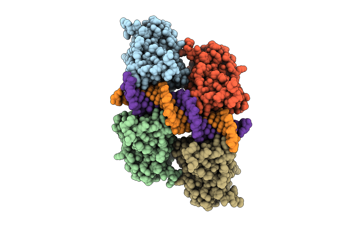
Crystal structure of p53 core domain in complex with DNA
Biological Source:
Source Organism:
Homo sapiens (Taxon ID: 9606)
(Taxon ID: )
Host Organism:
Method Details:
Experimental Method:
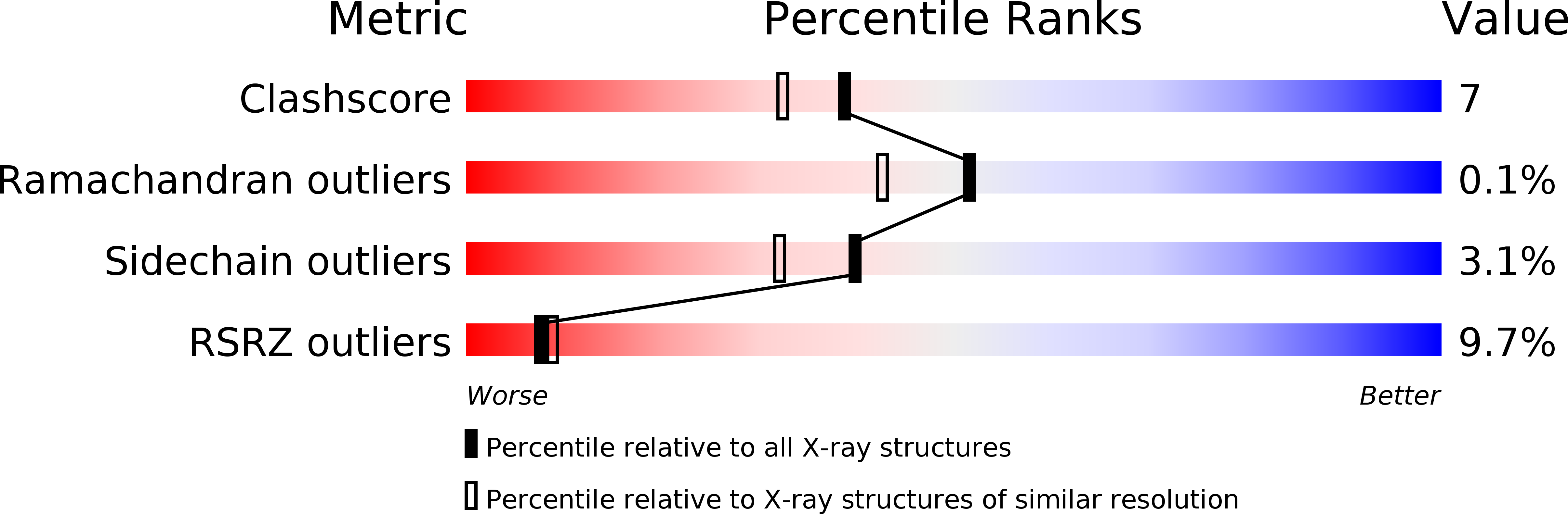
Resolution:
1.91 Å
R-Value Free:
0.22
R-Value Work:
0.18
R-Value Observed:
0.18
Space Group:
P 21 21 21
Macromolecular Entities
Polymer Type:polypeptide(L)
Molecule:Cellular tumor antigen p53
Gene (Uniprot):TP53
Chain IDs:A, B, C, D
Chain Length:200
Number of Molecules:4
Biological Source:Homo sapiens
Polymer Type:polydeoxyribonucleotide
Molecule:DNA (5'-D(*TP*CP*AP*CP*AP*AP*GP*TP*TP*AP*GP*AP*GP*AP*CP*AP*AP*GP*CP*CP*T)-3')
Chain IDs:E
Chain Length:21
Number of Molecules:1
Biological Source:
Polymer Type:polydeoxyribonucleotide
Molecule:DNA (5'-D(*AP*GP*GP*CP*TP*TP*GP*TP*CP*TP*CP*TP*AP*AP*CP*TP*TP*GP*TP*GP*A)-3')
Chain IDs:F
Chain Length:21
Number of Molecules:1
Biological Source:
Ligand Molecules
Primary Citation
Structure of p53 binding to the BAX response element reveals DNA unwinding and compression to accommodate base-pair insertion.
Nucleic Acids Res. 41 8368 8376 (2013)
PMID: 23836939 DOI: 10.1093/nar/gkt584

Abstact

The p53 core domain binds to response elements (REs) that contain two continuous half-sites as a cooperative tetramer, but how p53 recognizes discontinuous REs is not well understood. Here we describe the crystal structure of the p53 core domain bound to a naturally occurring RE located at the promoter of the Bcl-2-associated X protein (BAX) gene, which contains a one base-pair insertion between the two half-sites. Surprisingly, p53 forms a tetramer on the BAX-RE that is nearly identical to what has been reported on other REs with a 0-bp spacer. Each p53 dimer of the tetramer binds in register to a half-site and maintains the same protein-DNA interactions as previously observed, and the two dimers retain all the protein-protein contacts without undergoing rotation or translation. To accommodate the additional base pair, the DNA is deformed and partially disordered around the spacer region, resulting in an apparent unwinding and compression, such that the interactions between the dimers are maintained. Furthermore, DNA deformation within the p53-bound BAX-RE is confirmed in solution by site-directed spin labeling measurements. Our results provide a structural insight into the mechanism by which p53 binds to discontinuous sites with one base-pair spacer.

Legend

Protein

Chemical

Disease

Primary Citation of related structures