4HJ0 image
Deposition Date 2012-10-12
Release Date 2013-05-29
Last Version Date 2024-11-20
Entry Detail
PDB ID:
4HJ0
Keywords:
Title:
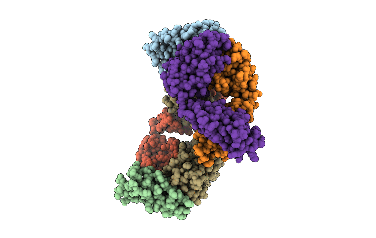
Crystal structure of the human GIPr ECD in complex with Gipg013 Fab at 3-A resolution
Biological Source:
Source Organism:
Homo sapiens (Taxon ID: 9606)
Host Organism:
Method Details:
Experimental Method:
Resolution:
3.00 Å
R-Value Free:
0.31
R-Value Work:
0.25
R-Value Observed:
0.26
Space Group:
P 1 21 1
Macromolecular Entities
Polymer Type:polypeptide(L)
Molecule:Gastric inhibitory polypeptide receptor
Gene (Uniprot):GIPR
Chain IDs:A, B
Chain Length:136
Number of Molecules:2
Biological Source:Homo sapiens
Polymer Type:polypeptide(L)
Molecule:Gipg013 Fab, Antagonizing antibody to the GIP Receptor, Heavy chain
Chain IDs:C (auth: P), F (auth: C)
Chain Length:227
Number of Molecules:2
Biological Source:Homo sapiens
Polymer Type:polypeptide(L)
Molecule:Gipg013 Fab, Antagonizing antibody to the GIP Receptor, Light chain
Chain IDs:D (auth: Q), E (auth: D)
Chain Length:215
Number of Molecules:2
Biological Source:Homo sapiens
Ligand Molecules
Primary Citation
Structural and Pharmacological Characterization of Novel Potent and Selective Monoclonal Antibody Antagonists of Glucose-dependent Insulinotropic Polypeptide Receptor.
J.Biol.Chem. 288 19760 19772 (2013)
PMID: 23689510 DOI: 10.1074/jbc.M112.426288

Abstact

Glucose-dependent insulinotropic polypeptide (GIP) is an endogenous hormonal factor (incretin) that, upon binding to its receptor (GIPr; a class B G-protein-coupled receptor), stimulates insulin secretion by beta cells in the pancreas. There has been a lack of potent inhibitors of the GIPr with prolonged in vivo exposure to support studies on GIP biology. Here we describe the generation of an antagonizing antibody to the GIPr, using phage and ribosome display libraries. Gipg013 is a specific competitive antagonist with equally high potencies to mouse, rat, dog, and human GIP receptors with a Ki of 7 nm for the human GIPr. Gipg013 antagonizes the GIP receptor and inhibits GIP-induced insulin secretion in vitro and in vivo. A crystal structure of Gipg013 Fab in complex with the human GIPr extracellular domain (ECD) shows that the antibody binds through a series of hydrogen bonds from the complementarity-determining regions of Gipg013 Fab to the N-terminal α-helix of GIPr ECD as well as to residues around its highly conserved glucagon receptor subfamily recognition fold. The antibody epitope overlaps with the GIP binding site on the GIPr ECD, ensuring competitive antagonism of the receptor. This well characterized antagonizing antibody to the GIPr will be useful as a tool to further understand the biological roles of GIP.

Legend

Protein

Chemical

Disease

Primary Citation of related structures