4HIP image
Deposition Date 2012-10-11
Release Date 2012-11-21
Last Version Date 2025-03-26
Entry Detail
PDB ID:
4HIP
Title:
Crystal structure of the Pseudomonas aeruginosa azurin, H126NO YOH109
Biological Source:
Source Organism:
Host Organism:
Method Details:
Experimental Method:
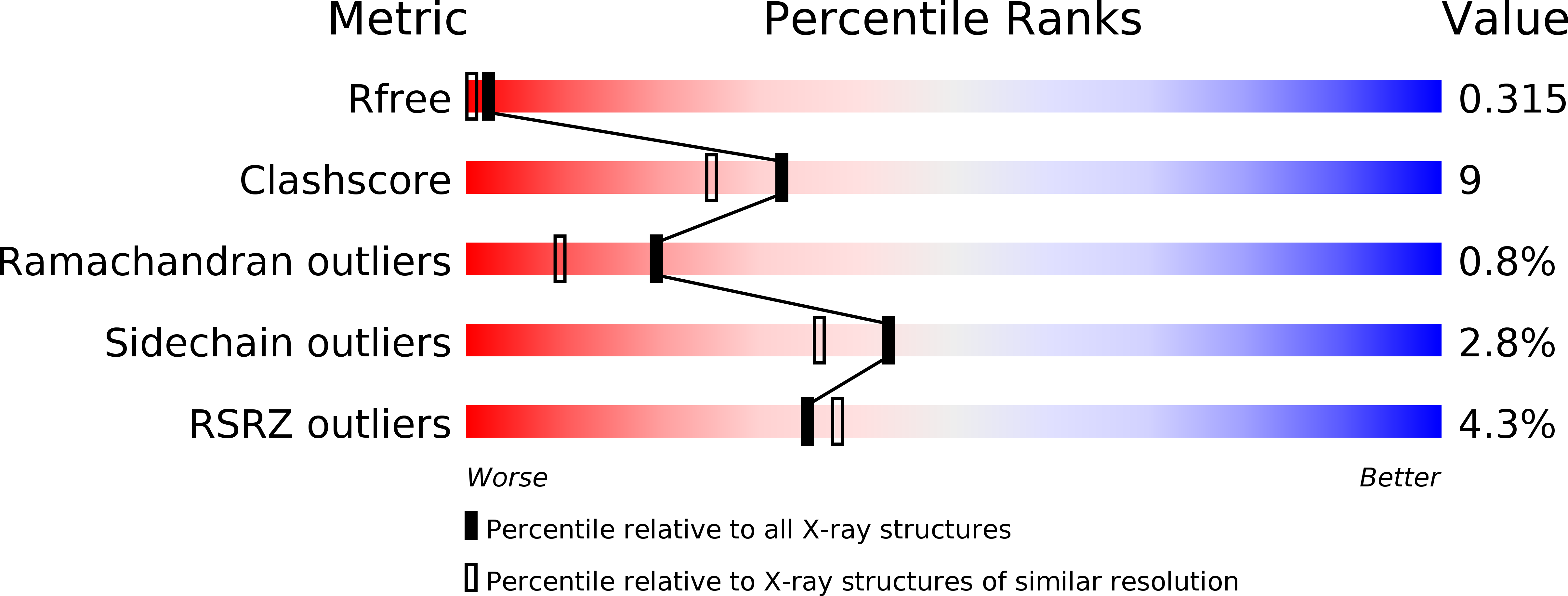
Resolution:
1.90 Å
R-Value Free:
0.31
R-Value Work:
0.24
R-Value Observed:
0.25
Space Group:
P 2 21 21
Macromolecular Entities
Polymer Type:polypeptide(L)
Molecule:Azurin
Gene (Uniprot):azu
Mutations:W68F,Y92F,H103Q,Y128F,K142NIY,T146H
Chain IDs:A, B
Chain Length:128
Number of Molecules:2
Biological Source:Pseudomonas aeruginosa
Modified Residue
Compound ID Chain ID Parent Comp ID Details 2D Image
NIY A TYR META-NITRO-TYROSINE
Ligand Molecules
Primary Citation
Electron Flow through Nitrotyrosinate in Pseudomonas aeruginosa Azurin.
J.Am.Chem.Soc. 135 11151 11158 (2013)
PMID: 23859602 DOI: 10.1021/ja403734n

Abstact

We have designed ruthenium-modified Pseudomonas aeruginosa azurins that incorporate 3-nitrotyrosine (NO2YOH) between Ru(2,2'-bipyridine)2(imidazole)(histidine) and Cu redox centers in electron transfer (ET) pathways. We investigated the structures and reactivities of three different systems: RuH107NO2YOH109, RuH124NO2YOH122, and RuH126NO2YOH122. RuH107NO2YOH109, unlabeled H124NO2YOH122, and unlabeled H126NO2YOH122 were structurally characterized. The pKa's of NO2YOH at positions 122 and 109 are 7.2 and 6.0, respectively. Reduction potentials of 3-nitrotyrosinate (NO2YO(-))-modified azurins were estimated from cyclic and differential pulse voltammetry data: oxidation of NO2YO(-)122 occurs near 1.1 versus NHE; oxidation of NO2YO(-)109 is near 1.2 V. Our analysis of transient optical spectroscopic experiments indicates that hopping via NO2YO(-) enhances Cu(I) oxidation rates over single-step ET by factors of 32 (RuH107NO2YO(-)109), 46 (RuH126NO2YO(-)122), and 13 (RuH124NO2YO(-)122).

Legend

Protein

Chemical

Disease

Primary Citation of related structures