Deposition Date
2012-10-11
Release Date
2013-10-16
Last Version Date
2024-02-28
Entry Detail
PDB ID:
4HI0
Keywords:
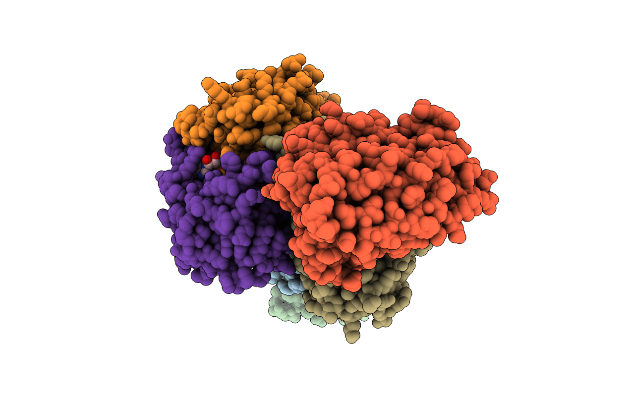
Title:
Crystal Structure of Helicobacter pylori Urease Accessory Protein UreF/H/G complex
Biological Source:
Source Organism(s):
Helicobacter pylori (Taxon ID: 85962)
Expression System(s):
Method Details:
Experimental Method:
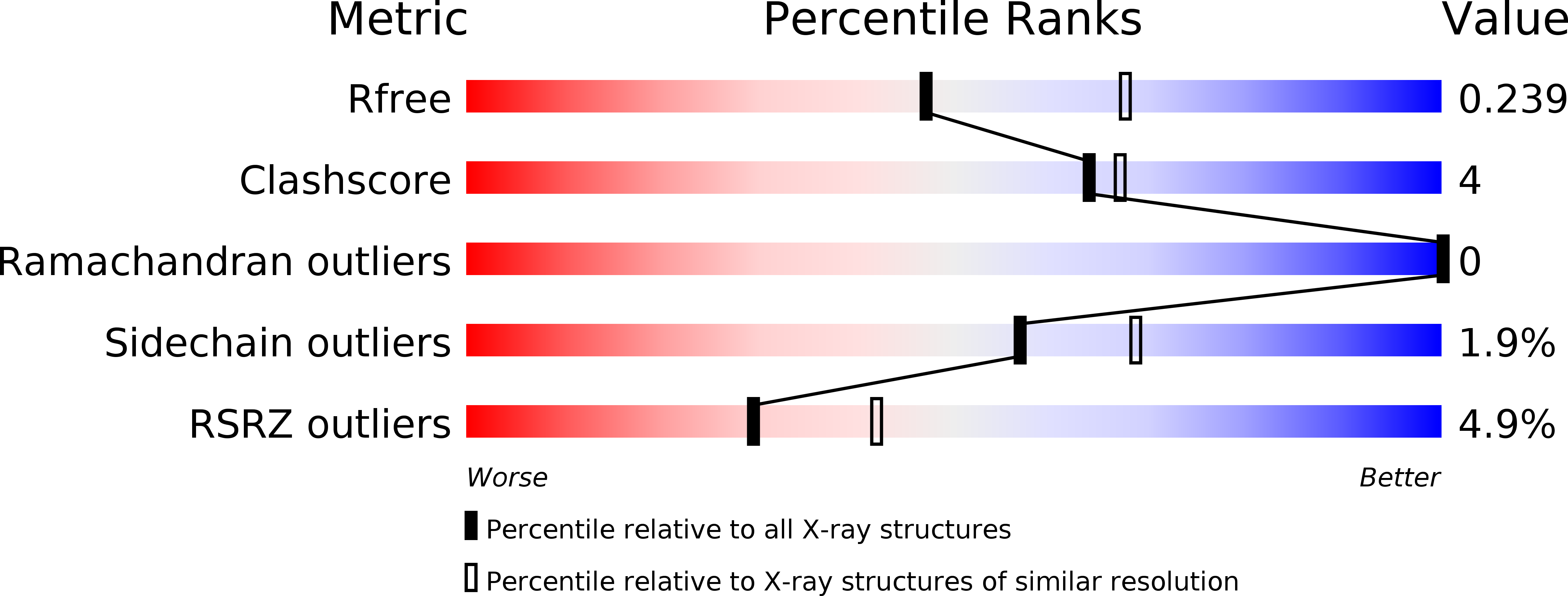
Resolution:
2.35 Å
R-Value Free:
0.24
R-Value Work:
0.19
R-Value Observed:
0.19
Space Group:
P 21 21 21