Deposition Date
2012-10-10
Release Date
2013-03-27
Last Version Date
2023-09-20
Entry Detail
PDB ID:
4HHL
Keywords:
Title:
High resolution crystal structure of Glucose Isomerase from Streptomyces sp. SK
Biological Source:
Source Organism(s):
Streptomyces sp. SK (Taxon ID: 253732)
Expression System(s):
Method Details:
Experimental Method:
Resolution:
1.73 Å
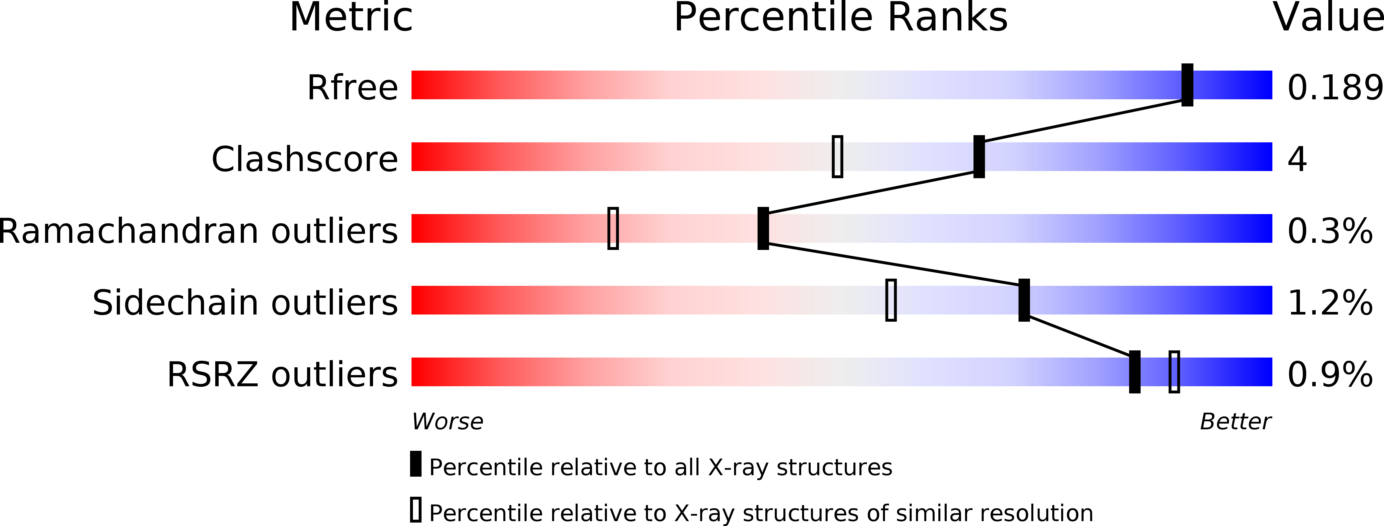
R-Value Free:
0.18
R-Value Work:
0.13
R-Value Observed:
0.13
Space Group:
C 1 2 1