4HF5 image
Deposition Date 2012-10-04
Release Date 2013-02-13
Last Version Date 2024-10-30
Entry Detail
PDB ID:
4HF5
Title:
Crystal structure of Fab 8F8 in complex a H2N2 influenza virus hemagglutinin
Biological Source:
Source Organism:
Influenza A virus (Taxon ID: 382813)
Homo sapiens (Taxon ID: 9606)
Host Organism:
Method Details:
Experimental Method:
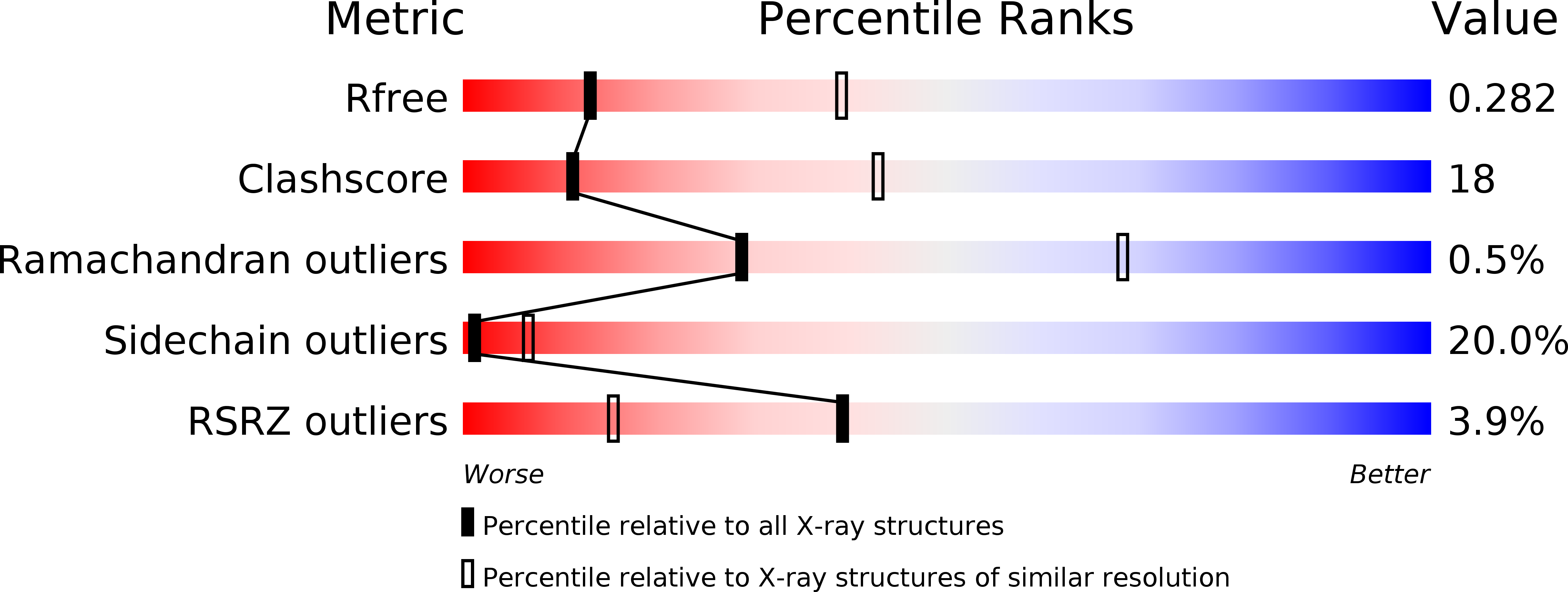
Resolution:
3.00 Å
R-Value Free:
0.28
R-Value Work:
0.22
R-Value Observed:
0.23
Space Group:
P 3 2 1
Macromolecular Entities
Structures with similar UniProt ID
Protein Blast
Polymer Type:polypeptide(L)
Molecule:Hemagglutinin HA1
Gene (Uniprot):HA
Chain IDs:A
Chain Length:327
Number of Molecules:1
Biological Source:Influenza A virus
Structures with similar UniProt ID
Protein Blast
Polymer Type:polypeptide(L)
Molecule:Hemagglutinin HA2
Gene (Uniprot):HA
Chain IDs:B
Chain Length:174
Number of Molecules:1
Biological Source:Influenza A virus
Polymer Type:polypeptide(L)
Molecule:Fab 8F8 heavy chain
Chain IDs:C (auth: H)
Chain Length:233
Number of Molecules:1
Biological Source:Homo sapiens
Polymer Type:polypeptide(L)
Molecule:Fab 8F8 light chain
Chain IDs:D (auth: L)
Chain Length:218
Number of Molecules:1
Biological Source:Homo sapiens
Modified Residue
Compound ID Chain ID Parent Comp ID Details 2D Image
ASN A ASN GLYCOSYLATION SITE
Ligand Molecules
Primary Citation
A recurring motif for antibody recognition of the receptor-binding site of influenza hemagglutinin.
Nat.Struct.Mol.Biol. 20 363 370 (2013)
PMID: 23396351 DOI: 10.1038/nsmb.2500

Abstact

Influenza virus hemagglutinin (HA) mediates receptor binding and viral entry during influenza infection. The development of receptor analogs as viral-entry blockers has not been successful, which suggests that sialic acid may not be an ideal scaffold to obtain broad, potent HA inhibitors. Here, we report crystal structures of Fab fragments from three human antibodies that neutralize the 1957 pandemic H2N2 influenza virus in complex with H2 HA. All three antibodies use an aromatic residue to plug a conserved cavity in the HA receptor-binding site. Each antibody interacts with the absolutely conserved HA1 Trp153 at the cavity base through π-π stacking with the signature Phe54 of two VH1-69-encoded antibodies or a tyrosine from HCDR3 in the other antibody. This highly conserved interaction can be used as a starting point to design inhibitors targeting this conserved hydrophobic pocket in influenza viruses.

Legend

Protein

Chemical

Disease

Primary Citation of related structures
Feedback Form
Name
Email
Institute
Feedback