4HE8 image
Deposition Date 2012-10-03
Release Date 2013-02-13
Last Version Date 2023-09-20
Entry Detail
PDB ID:
4HE8
Keywords:
Title:
Crystal structure of the membrane domain of respiratory complex I from Thermus thermophilus
Biological Source:
Source Organism:
Method Details:
Experimental Method:
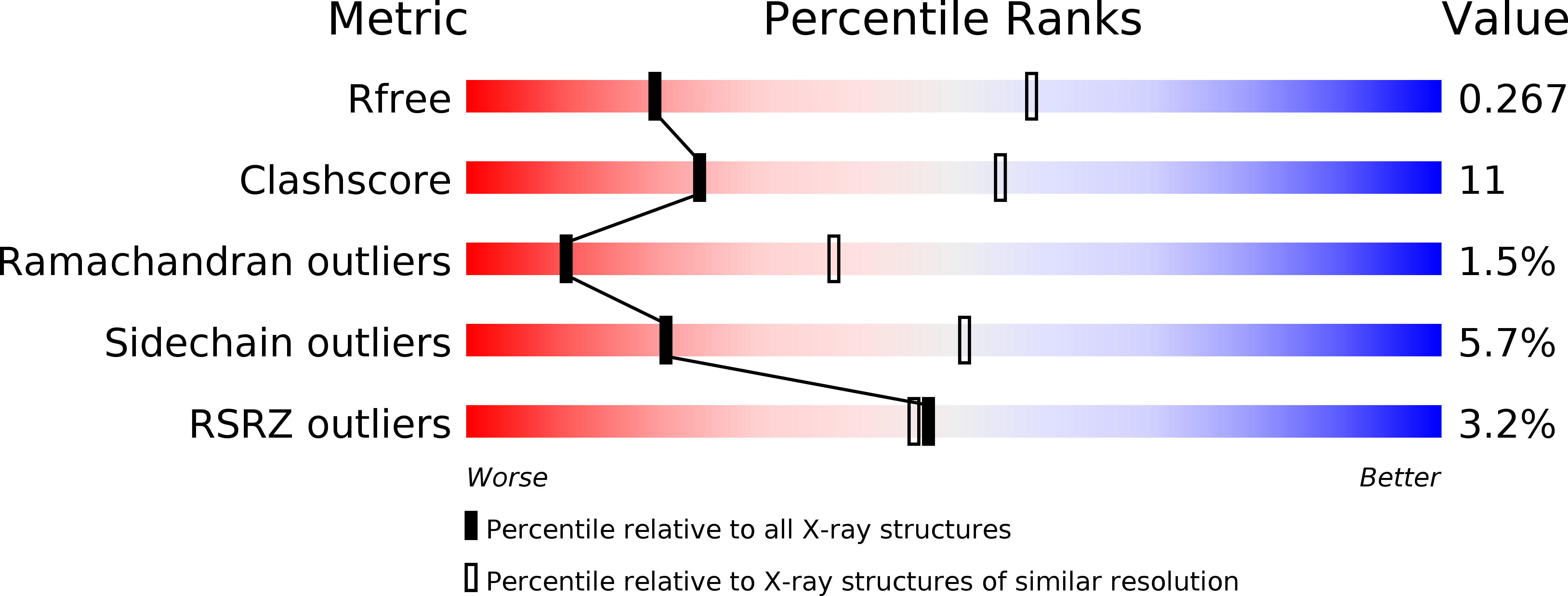
Resolution:
3.30 Å
R-Value Free:
0.26
R-Value Work:
0.20
R-Value Observed:
0.20
Space Group:
P 1
Macromolecular Entities
Polymer Type:polypeptide(L)
Molecule:NADH-quinone oxidoreductase subunit 7
Gene (Uniprot):nqo7
Chain IDs:A, H (auth: B)
Chain Length:119
Number of Molecules:2
Biological Source:Thermus thermophilus
Polymer Type:polypeptide(L)
Molecule:NADH-quinone oxidoreductase subunit 8
Gene (Uniprot):nqo8
Chain IDs:G (auth: H), N (auth: C)
Chain Length:365
Number of Molecules:2
Biological Source:Thermus thermophilus
Polymer Type:polypeptide(L)
Molecule:NADH-quinone oxidoreductase subunit 10
Gene (Uniprot):nqo10
Chain IDs:B (auth: J), I (auth: D)
Chain Length:176
Number of Molecules:2
Biological Source:Thermus thermophilus
Polymer Type:polypeptide(L)
Molecule:NADH-quinone oxidoreductase subunit 11
Gene (Uniprot):nqo11
Chain IDs:C (auth: K), J (auth: E)
Chain Length:95
Number of Molecules:2
Biological Source:Thermus thermophilus
Polymer Type:polypeptide(L)
Molecule:NADH-quinone oxidoreductase subunit 12
Gene (Uniprot):nqo12
Chain IDs:D (auth: L), K (auth: F)
Chain Length:606
Number of Molecules:2
Biological Source:Thermus thermophilus
Polymer Type:polypeptide(L)
Molecule:NADH-quinone oxidoreductase subunit 13
Gene (Uniprot):nqo13
Chain IDs:E (auth: M), L (auth: G)
Chain Length:469
Number of Molecules:2
Biological Source:Thermus thermophilus
Polymer Type:polypeptide(L)
Molecule:NADH-quinone oxidoreductase subunit 14
Gene (Uniprot):nqo14
Chain IDs:F (auth: N), M (auth: I)
Chain Length:427
Number of Molecules:2
Biological Source:Thermus thermophilus
Ligand Molecules
Primary Citation
Crystal structure of the entire respiratory complex I.
Nature 494 443 448 (2013)
PMID: 23417064 DOI: 10.1038/nature11871

Abstact

Complex I is the first and largest enzyme of the respiratory chain and has a central role in cellular energy production through the coupling of NADH:ubiquinone electron transfer to proton translocation. It is also implicated in many common human neurodegenerative diseases. Here, we report the first crystal structure of the entire, intact complex I (from Thermus thermophilus) at 3.3 Å resolution. The structure of the 536-kDa complex comprises 16 different subunits, with a total of 64 transmembrane helices and 9 iron-sulphur clusters. The core fold of subunit Nqo8 (ND1 in humans) is, unexpectedly, similar to a half-channel of the antiporter-like subunits. Small subunits nearby form a linked second half-channel, which completes the fourth proton-translocation pathway (present in addition to the channels in three antiporter-like subunits). The quinone-binding site is unusually long, narrow and enclosed. The quinone headgroup binds at the deep end of this chamber, near iron-sulphur cluster N2. Notably, the chamber is linked to the fourth channel by a 'funnel' of charged residues. The link continues over the entire membrane domain as a flexible central axis of charged and polar residues, and probably has a leading role in the propagation of conformational changes, aided by coupling elements. The structure suggests that a unique, out-of-the-membrane quinone-reaction chamber enables the redox energy to drive concerted long-range conformational changes in the four antiporter-like domains, resulting in translocation of four protons per cycle.

Legend

Protein

Chemical

Disease

Primary Citation of related structures