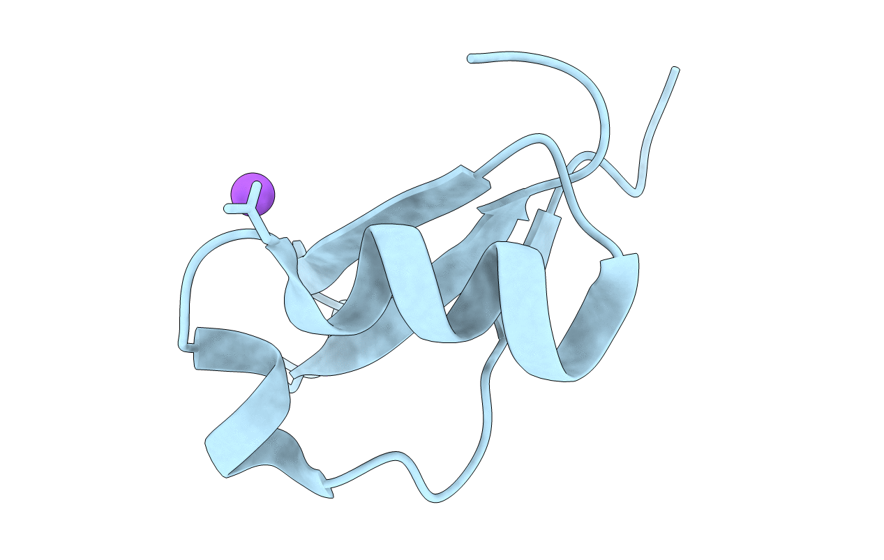
4HE7 image
Entry Detail
PDB ID:
4HE7
Keywords:
Title:
Crystal Structure of Brazzein
Biological Source:
Source Organism:
PDB Version:
Deposition Date:
2012-10-03
Release Date:
2013-03-27
Method Details:
Experimental Method:
Resolution:
1.80 Å
R-Value Free:
0.25
R-Value Work:
0.21
R-Value Observed:
0.21
Space Group:
I 41 2 2
Macromolecular Entities
Polymer Type:polypeptide(L)
Description:Defensin-like protein
Mutations:Q1E
Chain IDs:A
Chain Length:54
Number of Molecules:1
Biological Source:Pentadiplandra brazzeana
Modified Residue
Compound ID Chain ID Parent Comp ID Details 2D Image
PCA A GLN PYROGLUTAMIC ACID
Ligand Molecules
Primary Citation
The structure of brazzein, a sweet-tasting protein from the wild African plant Pentadiplandra brazzeana
Acta Crystallogr.,Sect.D 69 642 647 (2013)
PMID: 23519673 DOI: 10.1107/S0907444913001005

Abstact

Brazzein is the smallest sweet-tasting protein and was isolated from the wild African plant Pentadiplandra brazzeana. The brazzein molecule consists of 54 amino-acid residues and four disulfide bonds. Here, the first crystal structure of brazzein is reported at 1.8 Å resolution and is compared with previously reported solution structures. Despite the overall structural similarity, there are several remarkable differences between the crystal and solution structures both in their backbone folds and side-chain conformations. Firstly, there is an additional α-helix in the crystal structure. Secondly, the atomic r.m.s.d.s between the corresponding C(α)-atom pairs are as large as 2.0-2.2 Å between the crystal and solution structures. Thirdly, the crystal structure exhibits a molecular shape that is similar but not identical to the solution structures. The crystal structure of brazzein reported here will provide additional information and further insights into the intermolecular interaction of brazzein with the sweet-taste receptor.

Legend

Protein

Chemical

Disease

Primary Citation of related structures