Deposition Date
2012-10-03
Release Date
2013-05-29
Last Version Date
2024-11-06
Entry Detail
PDB ID:
4HE4
Keywords:
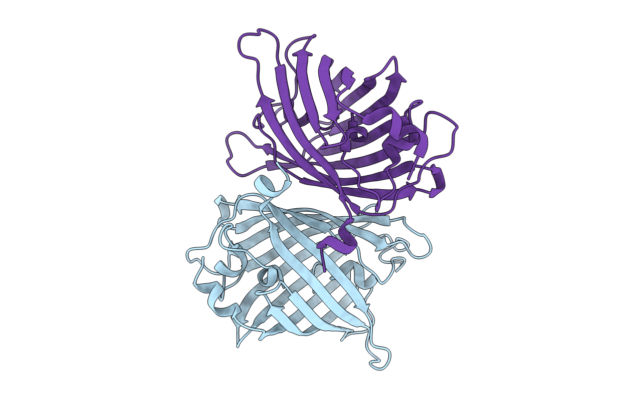
Title:
Crystal structure of the yellow fluorescent protein phiYFP (Phialidium sp.)
Biological Source:
Source Organism(s):
Phialidium sp. SL-2003 (Taxon ID: 258839)
Expression System(s):
Method Details:
Experimental Method:
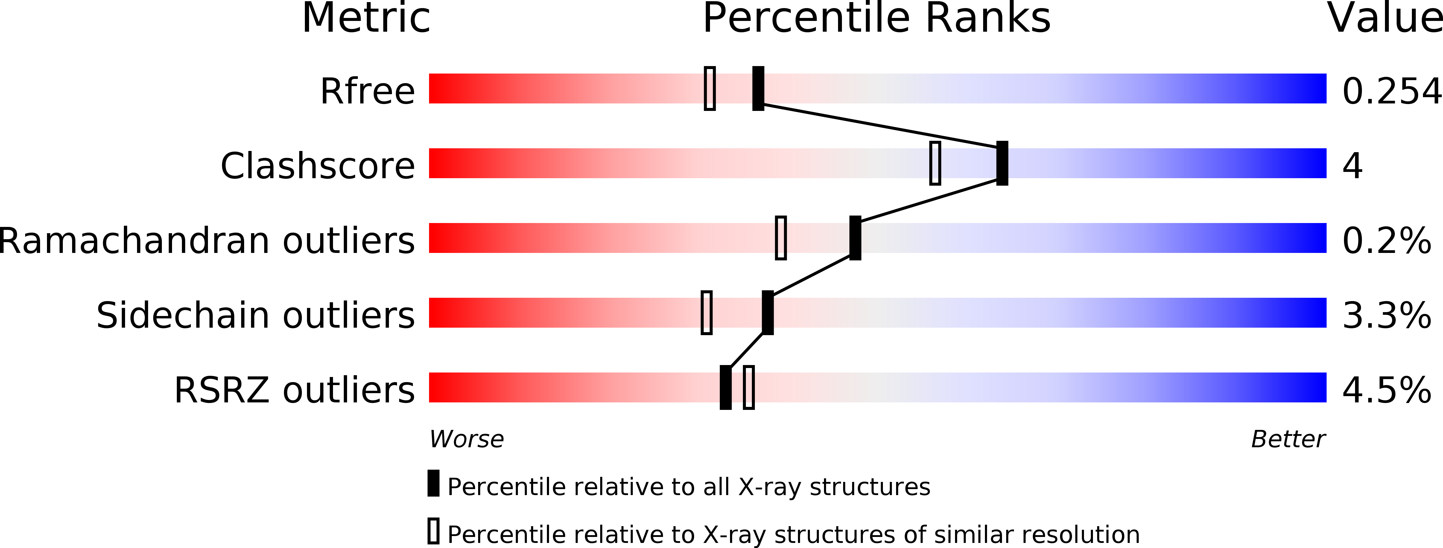
Resolution:
2.05 Å
R-Value Free:
0.25
R-Value Work:
0.19
R-Value Observed:
0.19
Space Group:
H 3 2