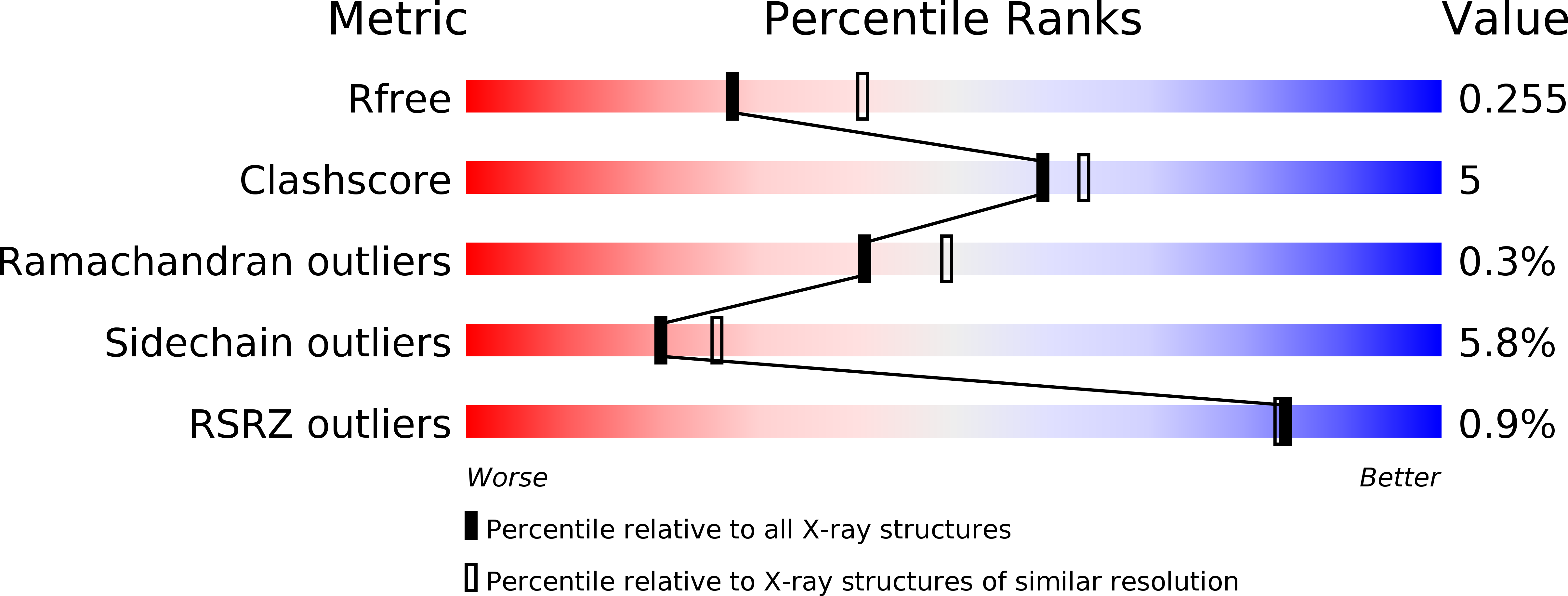
4HDI image
Deposition Date 2012-10-02
Release Date 2013-04-17
Last Version Date 2024-10-09
Entry Detail
PDB ID:
4HDI
Keywords:
Title:
Crystal Structure of 3E5 IgG3 FAB from mus musculus
Biological Source:
Source Organism:
Mus musculus (Taxon ID: 10090)
Host Organism:
Method Details:
Experimental Method:
Resolution:
2.45 Å
R-Value Free:
0.26
R-Value Work:
0.20
R-Value Observed:
0.21
Space Group:
P 21 21 21
Macromolecular Entities
Polymer Type:polypeptide(L)
Molecule:Ig heavy chain V region RF, Ig gamma-3 chain C region
Chain IDs:B (auth: H), D (auth: B)
Chain Length:220
Number of Molecules:2
Biological Source:Mus musculus
Polymer Type:polypeptide(L)
Molecule:Kappa light chain variable region, Anti-colorectal carcinoma light chain
Chain IDs:A (auth: L), C (auth: A)
Chain Length:219
Number of Molecules:2
Biological Source:Mus musculus
Primary Citation
Global structures of IgG isotypes expressing identical variable regions.
Mol.Immunol. 56 588 598 (2013)
PMID: 23911417 DOI: 10.1016/j.molimm.2013.06.006

Abstact

Until relatively recently the immunoglobulin molecule was viewed as composed of two independent domains comprised of the variable (V) and constant (C) regions. However, recent work has established that the C region mediates allosteric changes in the V region that can influence specificity and affinity. To further explore cross-domain interrelationship in murine IgG structure we carried out solution small angle X-ray scattering (SAXS) measurements for four V region identical IgG isotypes. SAXS analysis revealed elongated Y-shaped structures in solution with significantly different, isotype-dependent domain orientations. To further explore local C region effects on the V region, the IgG₃ Fab crystal structure from the same family was determined to 2.45 Å resolution. The IgG₃ Fab crystal structure differs from a closely related previously solved IgG1 Fab revealing significant structural differences, which may account for isotype-related specificity differences in V region identical Abs. Among the four murine isotypes, IgG₃ was the most different in solution with regards to overall structure as well as aggregate formation in solution suggesting that the greater apparent affinity of this isotype resulted from polyvalent complexes with enhanced avidity. Our results provide additional evidence that Ig V and C domains influence each other structurally and suggest that V region structure can have significant effects on overall Ig structure.

Legend

Protein

Chemical

Disease

Primary Citation of related structures