4HAC image
Deposition Date 2012-09-26
Release Date 2012-12-12
Last Version Date 2024-10-30
Entry Detail
PDB ID:
4HAC
Keywords:
Title:
Crystal Structure of the Mevalonate Kinase from an Archaeon Methanosarcina mazei
Biological Source:
Source Organism:
Host Organism:
Method Details:
Experimental Method:
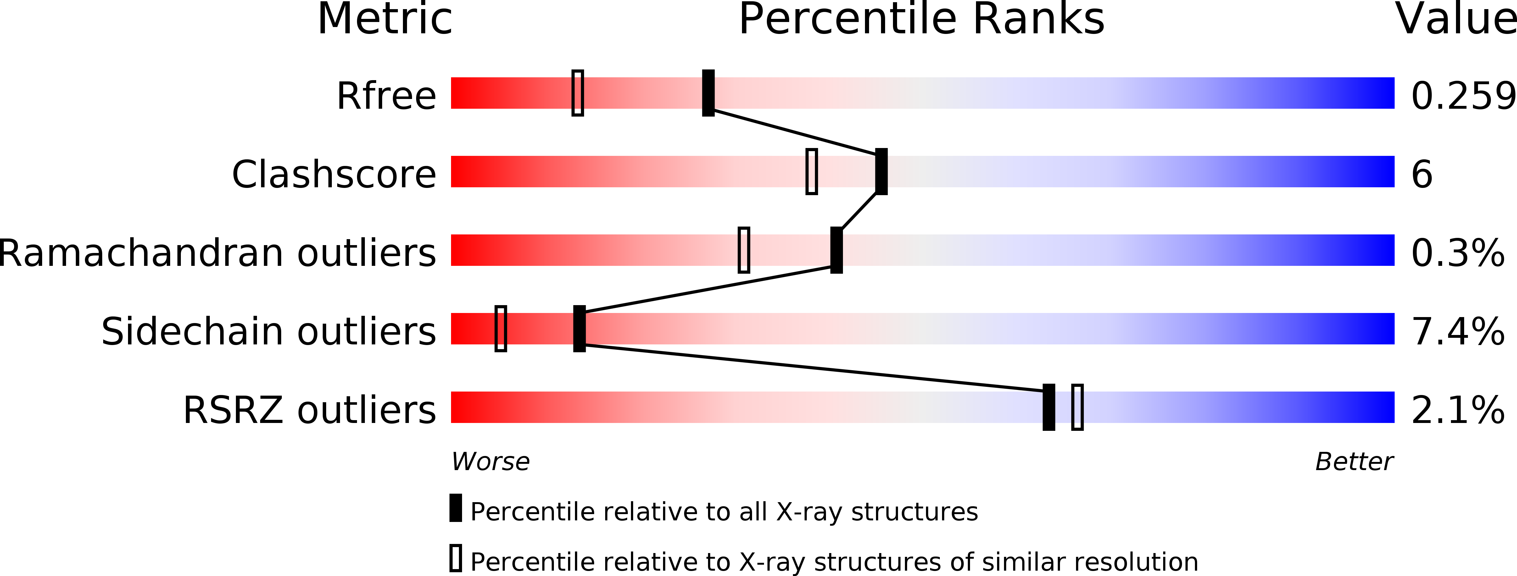
Resolution:
1.92 Å
R-Value Free:
0.26
R-Value Work:
0.21
R-Value Observed:
0.21
Space Group:
P 21 21 2
Macromolecular Entities
Structures with similar UniProt ID
Protein Blast
Polymer Type:polypeptide(L)
Molecule:Mevalonate kinase
Gene (Uniprot):mvk
Chain IDs:A
Chain Length:321
Number of Molecules:1
Biological Source:Methanosarcina mazei
Structures with similar UniProt ID
Protein Blast
Polymer Type:polypeptide(L)
Molecule:Mevalonate kinase
Gene (Uniprot):mvk
Chain IDs:B
Chain Length:321
Number of Molecules:1
Biological Source:Methanosarcina mazei
Modified Residue
Compound ID Chain ID Parent Comp ID Details 2D Image
MSE A MET SELENOMETHIONINE
Ligand Molecules
Primary Citation
Crystallization and preliminary X-ray diffraction analysis of mevalonate kinase from Methanosarcina mazei.
Acta Crystallogr.,Sect.F 68 1560 1563 (2012)
PMID: 23192048 DOI: 10.1107/S1744309112047070

Abstact

Mevalonate kinase (MVK), which plays an important role in catalysing the biosynthesis of isoprenoid compounds derived from the mevalonate pathway, transforms mevalonate to 5-phosphomevalonate using ATP as a cofactor. Mevalonate kinase from Methanosarcina mazei (MmMVK) was expressed in Escherichia coli, purified and crystallized for structural analysis. Diffraction-quality crystals of MmMVK were obtained by the vapour-diffusion method using 0.32 M MgCl2, 0.08 M bis-tris pH 5.5, 16%(w/v) PEG 3350. The crystals belonged to space group P2(1)2(1)2, with unit-cell parameters a=97.11, b=135.92, c=46.03 Å. Diffraction data were collected to 2.08 Å resolution.

Legend

Protein

Chemical

Disease

Primary Citation of related structures