4HAA image
Entry Detail
PDB ID:
4HAA
Keywords:
Title:
Structure of Ribonuclease Binase Glu43Ala/Phe81Ala Mutant
Biological Source:
Source Organism:
Host Organism:
PDB Version:
Deposition Date:
2012-09-26
Release Date:
2012-10-17
Method Details:
Experimental Method:
Resolution:
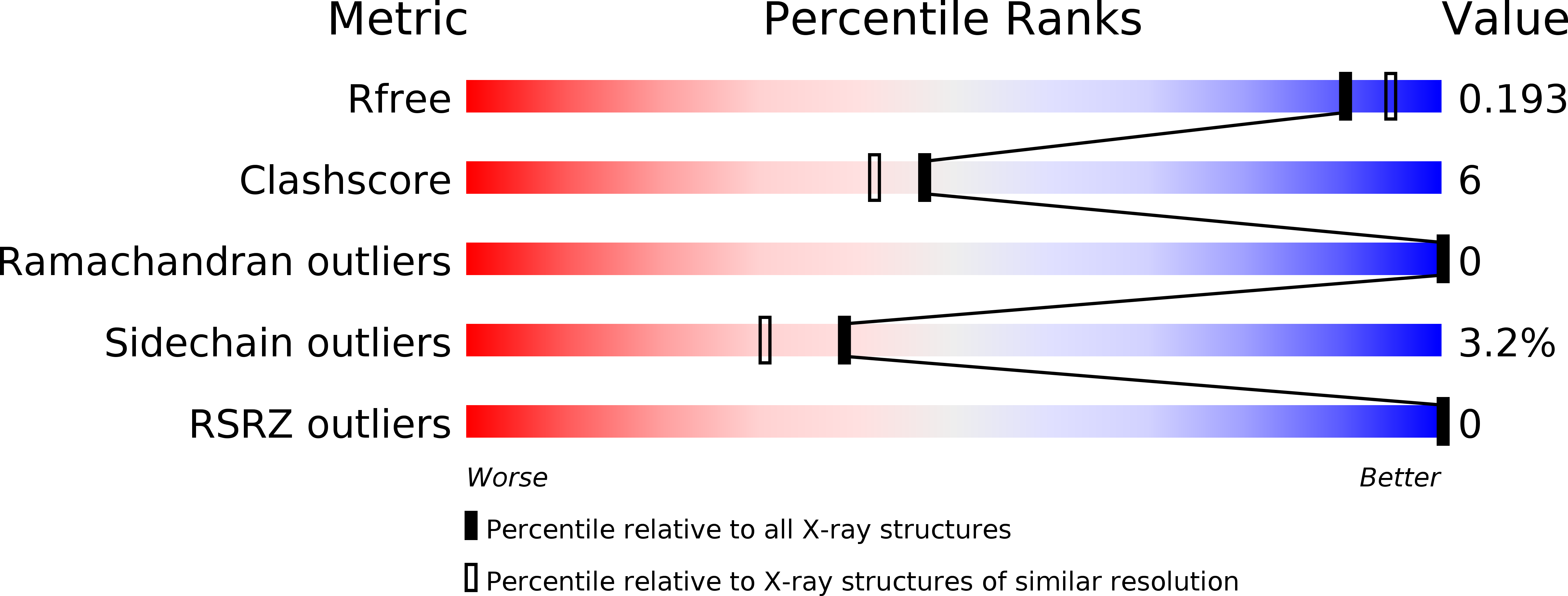
1.90 Å
R-Value Free:
0.20
R-Value Work:
0.17
R-Value Observed:
0.17
Space Group:
P 43
Macromolecular Entities
Polymer Type:polypeptide(L)
Description:Ribonuclease
Mutations:E43A/F81A
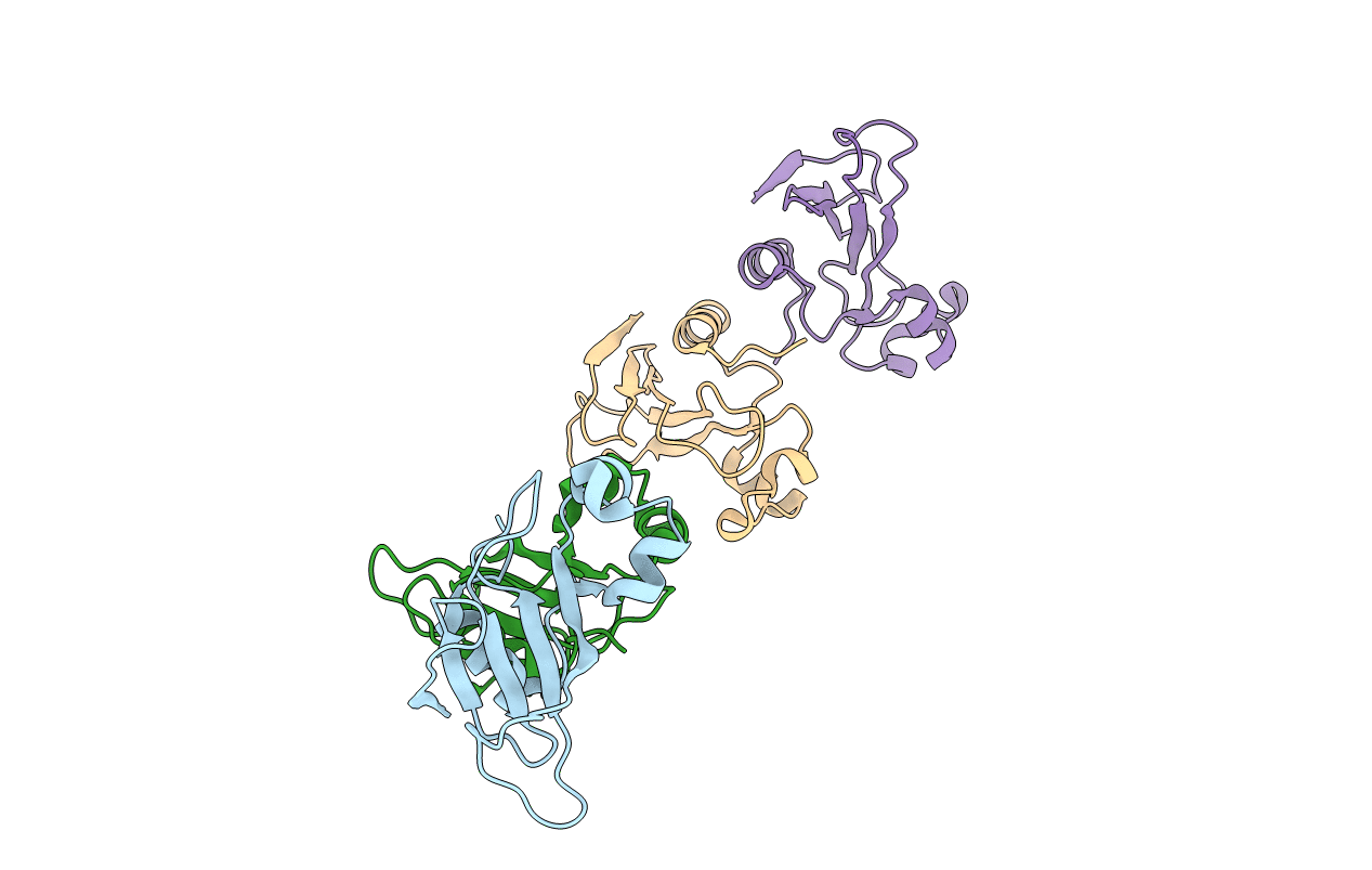
Chain IDs:A, B, C, D
Chain Length:109
Number of Molecules:4
Biological Source:Bacillus intermedius
Primary Citation
Structure and functional studies of the ribonuclease binase Glu43Ala/Phe81Ala mutant.
Acta Crystallogr.,Sect.D 69 991 996 (2013)
PMID: 23695243 DOI: 10.1107/S0907444913004046

Abstact

Ribonuclease from Bacillus intermedius (binase) is a small basic protein with antitumour activity. The three-dimensional structure of the binase mutant form Glu43Ala/Phe81Ala was determined at 1.98 Å resolution and its functional properties, such as the kinetic parameters characterizing the hydrolysis of polyinosinic acid and cytotoxicity towards Kasumi-1 cells, were investigated. In all crystal structures of binase studied previously the characteristic dimer is present, with the active site of one subunit being blocked owing to interactions within the dimer. In contrast to this, the new mutant form is not dimeric in the crystal. The catalytic efficiency of the mutant form is increased 1.7-fold and its cytotoxic properties are enhanced compared with the wild-type enzyme.

Legend

Protein

Chemical

Disease

Primary Citation of related structures