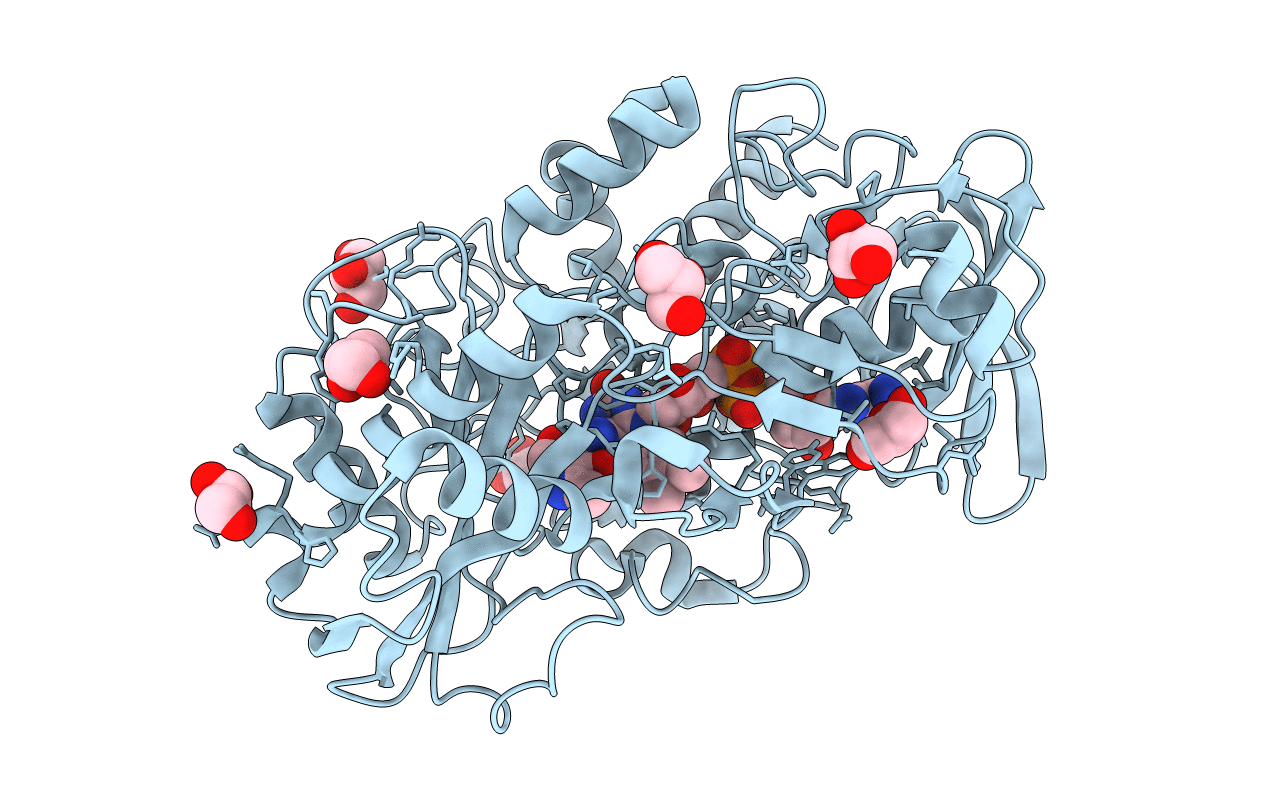
4HA6 image
Entry Detail
PDB ID:
4HA6
Keywords:
Title:
Crystal structure of pyridoxine 4-oxidase - pyridoxamine complex
Biological Source:
Source Organism:
Host Organism:
PDB Version:
Deposition Date:
2012-09-25
Release Date:
2013-06-05
Method Details:
Experimental Method:
Resolution:
2.10 Å
R-Value Free:
0.23
R-Value Work:
0.18
R-Value Observed:
0.18
Space Group:
P 21 21 21
Macromolecular Entities
Polymer Type:polypeptide(L)
Description:Pyridoxine 4-oxidase
Chain IDs:A
Chain Length:508
Number of Molecules:1
Biological Source:Mesorhizobium loti
Modified Residue
Compound ID Chain ID Parent Comp ID Details 2D Image
CME A CYS S,S-(2-HYDROXYETHYL)THIOCYSTEINE
Primary Citation
Crystal structure of pyridoxine 4-oxidase from Mesorhizobium loti.
Biochim.Biophys.Acta 1834 953 963 (2013)
PMID: 23501672 DOI: 10.1016/j.bbapap.2013.03.004

Abstact

Pyridoxine 4-oxidase (PNOX) from Mesorhizobium loti is a monomeric glucose-methanol-choline (GMC) oxidoreductase family enzyme, catalyzes FAD-dependent oxidation of pyridoxine (PN) into pyridoxal, and is the first enzyme in pathway I for the degradation of PN. The tertiary structures of PNOX with a C-terminal His6-tag and PNOX-pyridoxamine (PM) complex were determined at 2.2Å and at 2.1Å resolutions, respectively. The overall structure consisted of FAD-binding and substrate-binding domains. In the active site, His460, His462, and Pro504 were located on the re-face of the isoalloxazine ring of FAD. PM binds to the active site through several hydrogen bonds. The side chains of His462 and His460 are located at 2.7 and 3.1Å from the N4' atom of PM. The activities of His460Ala and His462Ala mutant PNOXs were very low, and 460Ala/His462Ala double mutant PNOX exhibited no activity. His462 may act as a general base for the abstraction of a proton from the 4'-hydroxyl of PN. His460 may play a role in the binding and positioning of PN. The C4' atom in PM is located at 3.2Å, and the hydride ion from the C4' atom may be transferred to the N5 atom of the isoalloxazine ring. The comparison of active site residues in GMC oxidoreductase shows that Pro504 in PNOX corresponds to Asn or His of the conserved His-Asn or His-His pair in other GMC oxidoreductases. The function of the novel proline residue was discussed.

Legend

Protein

Chemical

Disease

Primary Citation of related structures