Deposition Date
2012-09-20
Release Date
2012-10-03
Last Version Date
2023-09-20
Entry Detail
PDB ID:
4H7A
Keywords:
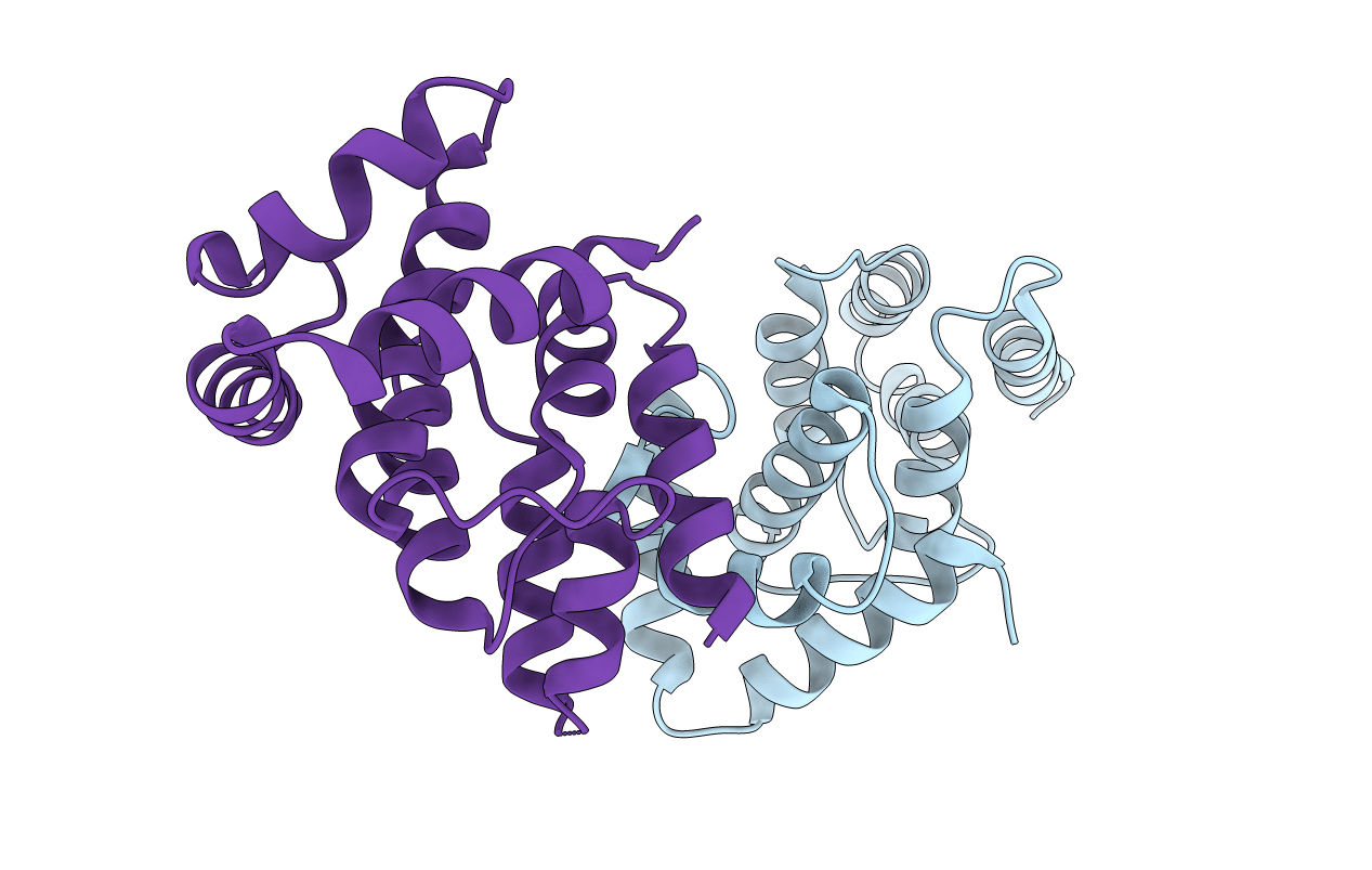
Title:
Crystal structure of CasB from Thermus thermophilus
Biological Source:
Source Organism(s):
Thermus thermophilus (Taxon ID: 300852)
Expression System(s):
Method Details:
Experimental Method:
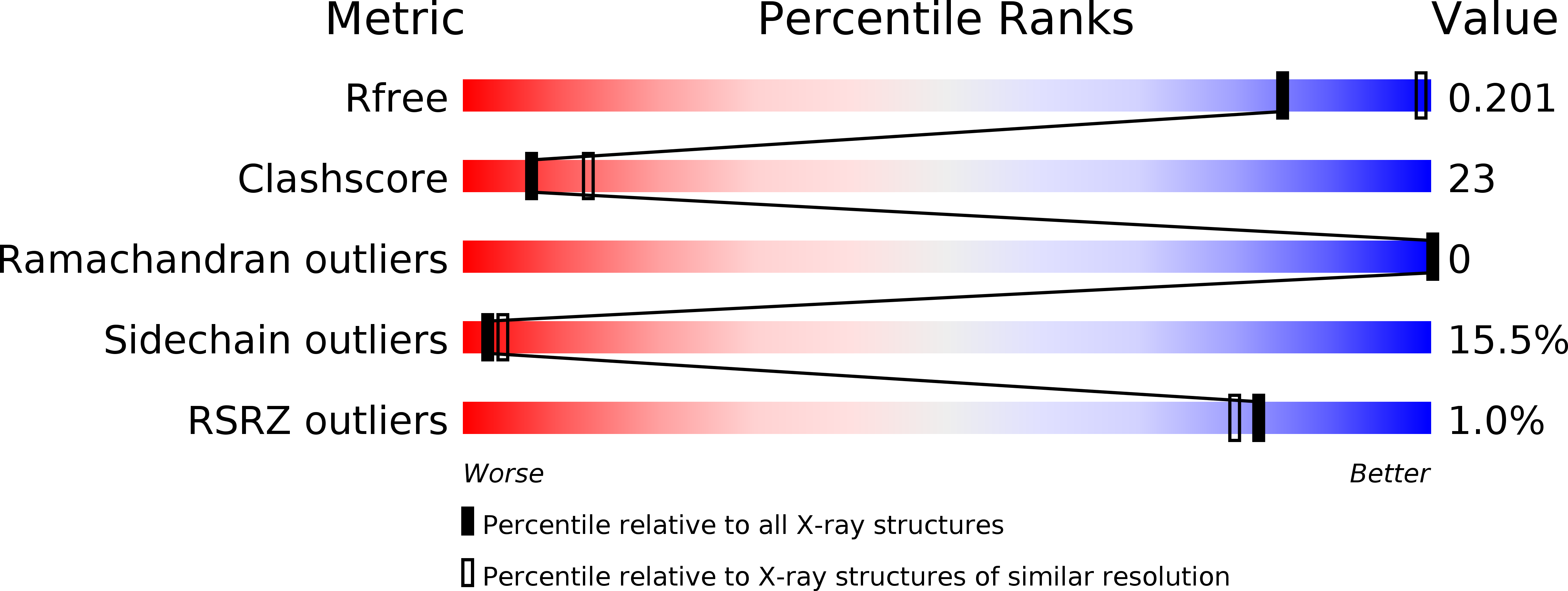
Resolution:
2.60 Å
R-Value Free:
0.22
R-Value Work:
0.17
R-Value Observed:
0.17
Space Group:
P 43 21 2